
|
Fig. 1
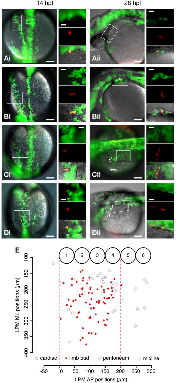
A single-cell-resolution fate map of the pectoral fin-field in the zebrafish LPM. (Ai) Dorsal view of 14hpf embryo previously injected with Kaede RNA in a single blastomere. Box shows a single cell photo-converted to express red fluorescence. Insets show the boxed region at higher magnification of green Kaede fluorescence (upper), red photo-converted cell (middle) and overlaid fluorescence with differential interference contrast microscopy (bottom). (Aii) Same embryo in lateral view at 28hpf. Box shows resultant clone in cardiac region; insets are as in Ai. (Bi,Bii) Fin bud clone. (Ci,Cii) Peritoneum clone. (Di,Dii) Midline clone. Scale bars: 100µm in overviews; 10µm in insets. (E) Fate map of LPM cell fates at 14hpf. x-axis: AP position relative to somite borders. y-axis: ML position from midline of embryo. Numbered circles denote somite positions. Red dashed lines delineate the fin-field at 14hpf. dc, duct of Cuvier; ML, mediolateral; o, otic vesicle.