
IMAGE
Fig. 3
- ID
- ZDB-IMAGE-160205-67
- Publication
- Smith et al., 2015 - Analysis of Zebrafish Larvae Skeletal Muscle Integrity with Evans Blue Dye
- All Figures
- Figures for Smith et al., 2015
Image
|
Figure Caption
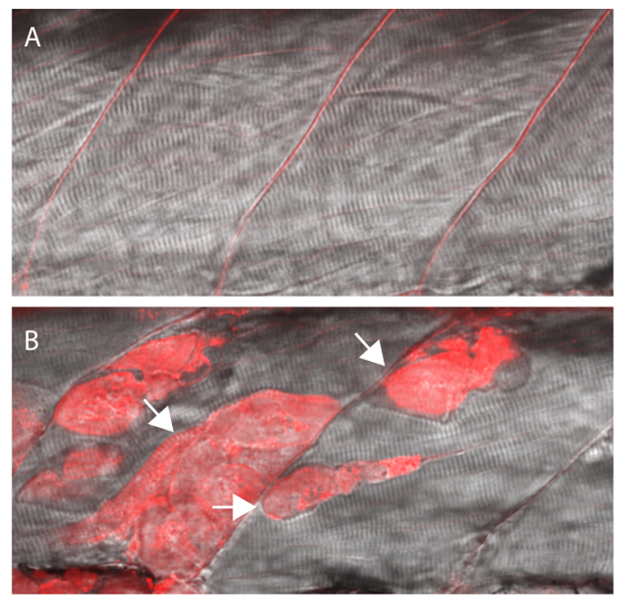
Fig. 3
EBD will be taken up by fibers with damaged membranes. (A) Wildtype siblings showing no EBD fluorescence in muscle fibers. (B) Sapje homozygous mutant with EBD fluorescence within multiple muscle fibers (arrows). All larvae were injected with the EBD injection mix and analyzed after a 4 hr incubation period at 3 dpf. Siblings and mutants were sorted by muscle fiber detachment prior to CCV injection.
Figure Data
Acknowledgments
This image is the copyrighted work of the attributed author or publisher, and
ZFIN has permission only to display this image to its users.
Additional permissions should be obtained from the applicable author or publisher of the image.
Full text @ J. Vis. Exp.