
|
Fig. S2
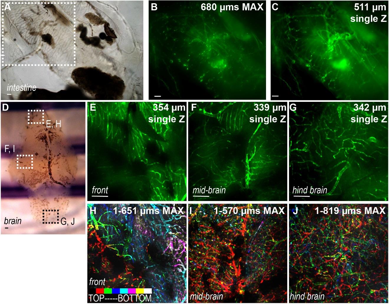
Whole animal clearing clears internal organs. (A,B,C) Intestine dissected from PACT-cleared whole Tg(Flk1:eGFP) animal is clear (A) and blood vessels can be imaged with an epiflourescent non-confocal microscope (B,C). (B) shows a 680 µm max projection, while (C) shows a single plane at a depth of 511 µm. (D) Brain dissected from PACT-cleared whole Tg(Flk1:eGFP) animal. (E,F,G) Single planes from indicated regions of the brain boxed in (D). (H,I,J) Depth-coded maximum projection images of brain vasculature. Scale bars are 100 µm. Single Z frames were exported and gamma adjusted in FIJI/ImageJ for increased visibility, with all gamma adjustments applied uniformly across images from either top or bottom stack.