
|
Fig. S1
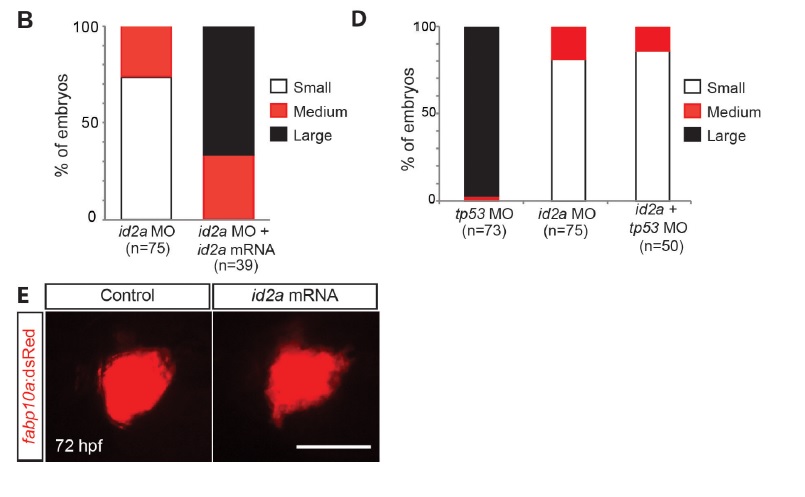
id2a mRNA, but not tp53 MO, injection rescues the reduced liver size in id2a MO-injected embryos. (A, B) Epifluorescence images showing fabp10a:dsRed expression (red) in control, id2a MO-injected, and id2a MO + mRNA-co-injected embryos (A) and their quantification (B). Although the liver of the co-injected embryos was still smaller than the control liver, it was much larger than the liver of the single MO-injected embryos, indicating a partial rescue of the liver size defect exhibited in id2a MO-injected embryos. For quantification, embryos were divided into three groups based on liver size: small, medium, and large. The id2a MO-injected liver size shown in A was considered as small; the liver size of the co-injected embryo shown in A was considered as large. Dorsal view, anterior to the left. (C) Bright-field and epifluorescence images showing the overall morphology of embryos and fabp10a:dsRed expression (red), respectively, in control, tp53 MO-injected, id2a MO-injected, and id2a MO + tp53 MO-co-injected embryos. Liver size as well as eye and head size (arrows) in id2a MO-injected embryos was similar to that in embryos co-injected withid2a and tp53 MOs. (D) Quantification of the results in C. For quantification, the liver size of the control embryo shown in C was considered as large and the liver size of the id2a MO-injected embryo shown in C was considered as small. Lateral view, anterior to the left. (E) Epifluorescence images showing the fabp10a:dsRed expression (red) in control and id2a mRNA-injected embryos at 72 hpf. Liver size in id2a-mRNA injected embryos was similar to that of control embryos. Lateral view, anterior to the left. Scale bars: 100 µm.
Reprinted from Mechanisms of Development, 138 Pt 3, Khaliq, M., Choi, T.Y., So, J., Shin, D., Id2a is required for hepatic outgrowth during liver development in zebrafish, 399-414, Copyright (2015) with permission from Elsevier. Full text @ Mech. Dev.