
Fig. 4
- ID
- ZDB-IMAGE-151228-14
- Genes
- Antibodies
- Publication
- Mendieta-Serrano et al., 2015 - Spatial and temporal expression of zebrafish glutathione peroxidase 4 a and b genes during early embryo development
- All Figures
- Figures for Mendieta-Serrano et al., 2015
|
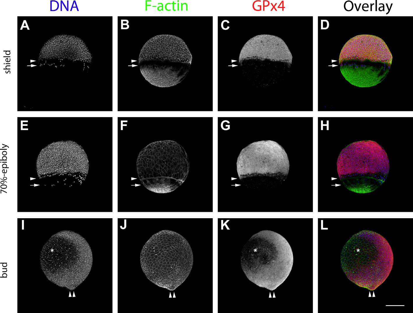
Fig. 4
GPx4 immunofluorescence localization patterns in zebrafish embryos at shield, 70%-epiboly and bud stages as detected by laser confocal microscopy. Shield stage embryos (A, B, C and D). 70%-epiboly (E, F, G and H). Bud stage embryos (I, J, K and L). Hoechst-stained embryos (A, E and I). F-actin, phalloidin Alexa 488-stained embryos (B, F and J). GPx4b immunolocalization (C, G and K). Left side views are shown. Dorsal side is to the right. Arrowheads indicate the epiboly migration front of the enveloping layer. Arrows indicate the position of the yolk nuclei epiboly migration front. Double arrowheads indicate tail bud position. *, evacuation zone. Scale bar 250 µm.
Reprinted from Gene expression patterns : GEP, 19(1-2), Mendieta-Serrano, M.A., Schnabel-Peraza, D., Lomelí, H., Salas-Vidal, E., Spatial and temporal expression of zebrafish glutathione peroxidase 4 a and b genes during early embryo development, 98-107, Copyright (2015) with permission from Elsevier. Full text @ Gene Expr. Patterns