
Image
|
Figure Caption
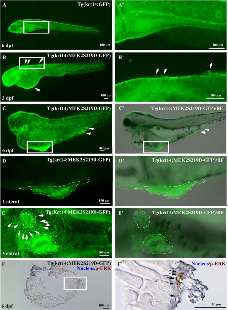
Fig. 4
Skin tumor formation in Tg(krt14: MEK2S219D-GFP) zebrafish. Skin cells proliferated and formed papilla from 3dfp (b, B′, white arrow) on the yolk skin in Tg(krt14: MEK2S219D-GFP) embryos to form a skin tumor at 6 dpf (c, and C′; lateral view, d and D′, white-dots area; ventral view, e and E′, white-dots area). Tg(krt14:GFP) embryos had developed normal skin cells at 6 dpf (a, A′). An immunohistochemical experiment was used to detect ERK phosphorylation in skin tumors. p-ERK was detected by a p-ERK monoclonal antibody and visualized by DAB (brown). Nuclei were counterstained with hematoxylin (blue) (f, F′)
Figure Data
Acknowledgments
This image is the copyrighted work of the attributed author or publisher, and
ZFIN has permission only to display this image to its users.
Additional permissions should be obtained from the applicable author or publisher of the image.
Full text @ J. Biomed. Sci.