
|
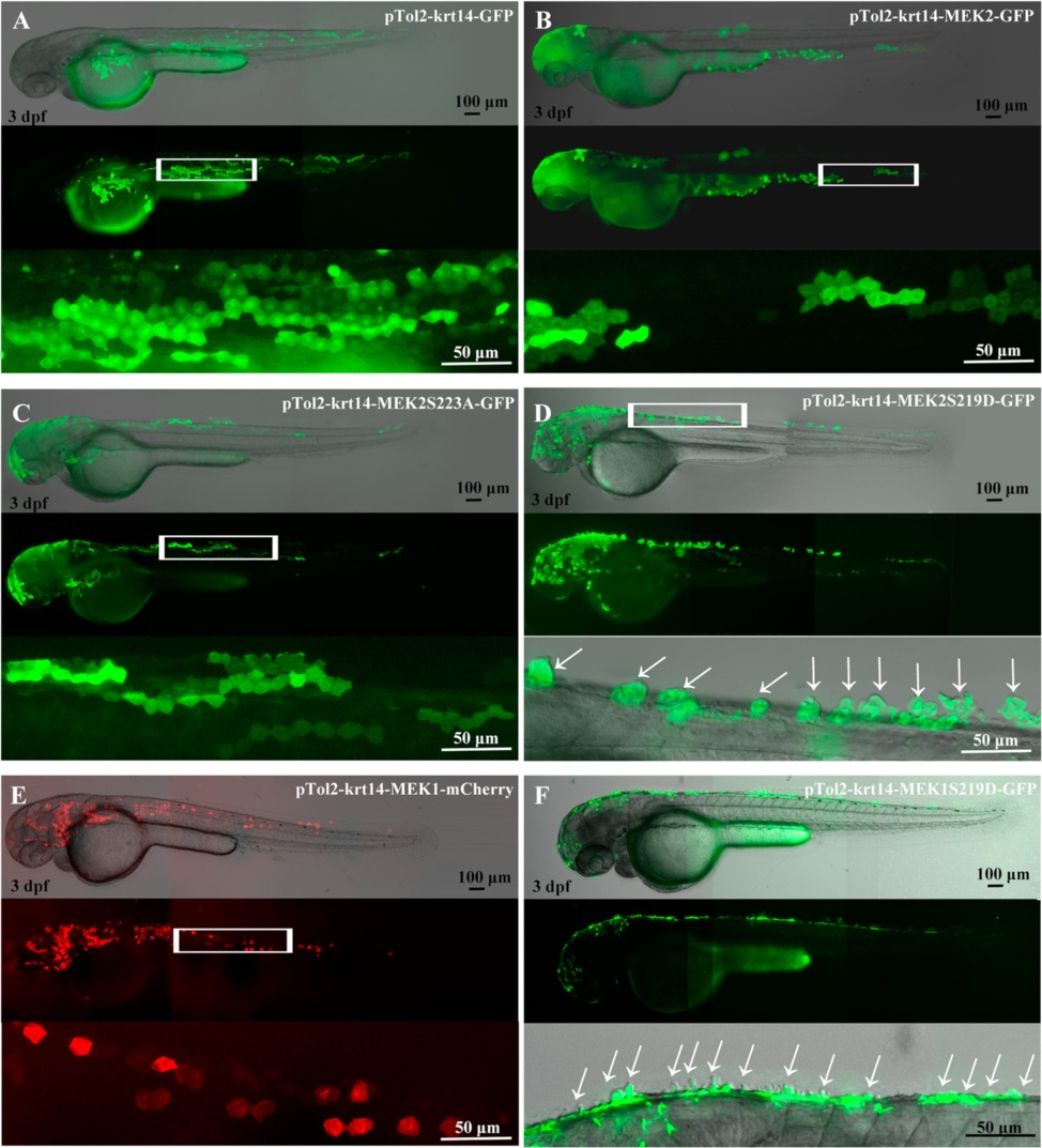
Fig. 3
Transient expressions of MEK1 and MEK2 driven by the krt14 promoter induced papillae formation in skin cells. Lateral view of pTol2-krt14-GFP (a), pTol2-krt14-MEK2-GFP (b), pTol2-MEK2S223A-GFP (c), pTol2-krt14-MEK2S219D-GFP (d), pTol2-krt14-MEK1-mCherry (e), and pTol2-krt14-MEK1S219D-GFP (f) plasmids, which were microinjected into 1-cell stage of zebrafish embryos and visualized at 3 days post-fertilization (dpf). The arrow indicates skin cell papillae and budding in the upper epidermis. The upper panel is a fluorescent image. The middle panel is merged fluorescent and bright-field images. The lower panel is an enlargement of the white box in the middle panel