
|
Fig. 3
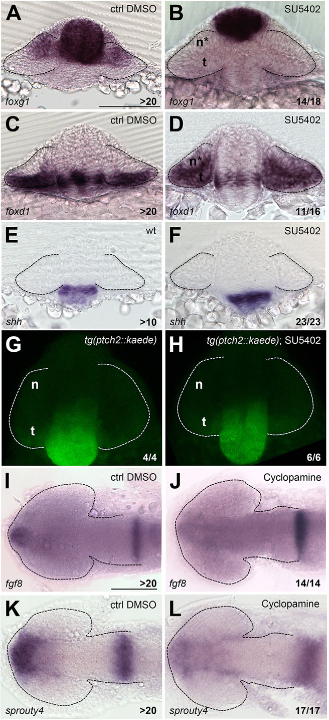
Lack of Fgf activity alters NT patterning independently of Shh activity. (A-L) Expression of foxg1 (A,B), foxd1 (C,D), shh (E,F), Kaede (G,H), fgf8 (I,J) and sprouty4 (K,L) in the conditions specified in the panels. A-H are frontal views; I-L are dorsal views with anterior to the left. All embryos are at 10-12ss. Scale bars: 100µm. Numbers in the bottom-right of each panel indicate the number of embryos with the phenotype shown out of the total number of embryos analysed. n, nasal; t, temporal; n*, defective nasal domain. Dashed lines outline the forebrain (dorsal views) or the optic vesicles (frontal views).