
|
Fig. 4
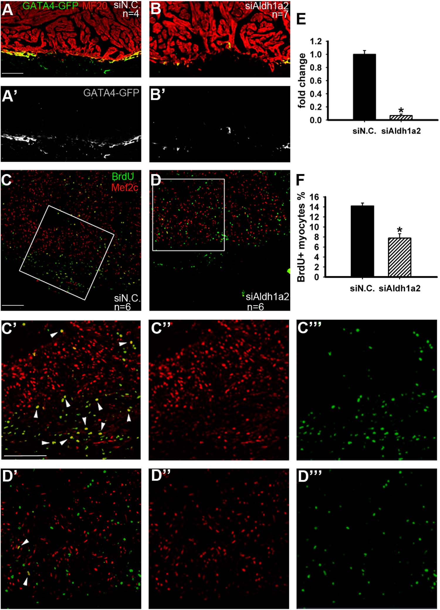
Nanoparticle-aided suppression of Aldh1a2 impedes cardiac regeneration. (A, B) siAldh1a2 inhibited Gata4-GFP cardiomyocytes in the injured areas (B, B′) compared with negative (siN.C.) control treatment (A, A′). MF20 marks the myocardium. (C, D) siAldh1a2 diminished the numbers of BrdU+/Mef2C+ proliferating myocytes (D, D′) compared with siN.C. treatment (C, C′) at 14 dpa. (E) Statistics of Gata4-GFP cardiomyocytes from A, B. (F) Statistics of Mef2C+/BrdU+ proliferating myocytes from C, D. (C′, C′′, C′′′) High-magnification images of the squared image in panel C showing Mef2C+/BrdU+ colocalization (C′), red fluorescent channel for Mef2C (C′′) and green fluorescent channel for BrdU (C′′′). (D′, D′′, D′′′) High-magnification images of the squared image in panel D showing Mef2C+/BrdU+ colocalization (D′), red fluorescent channel for Mef2C (D′′) and green fluorescent channel for BrdU (D′′′). *P<0.05; scale bars for A–D, 100 µm.
Reprinted from Developmental Biology, 406(2), Diao, J., Wang, H., Chang, N., Zhou, X.H., Zhu, X., Wang, J., Xiong, J.W., PEG-PLA Nanoparticles facilitate siRNA knockdown in adult zebrafish heart, 196-202, Copyright (2015) with permission from Elsevier. Full text @ Dev. Biol.