
Image
|
Figure Caption
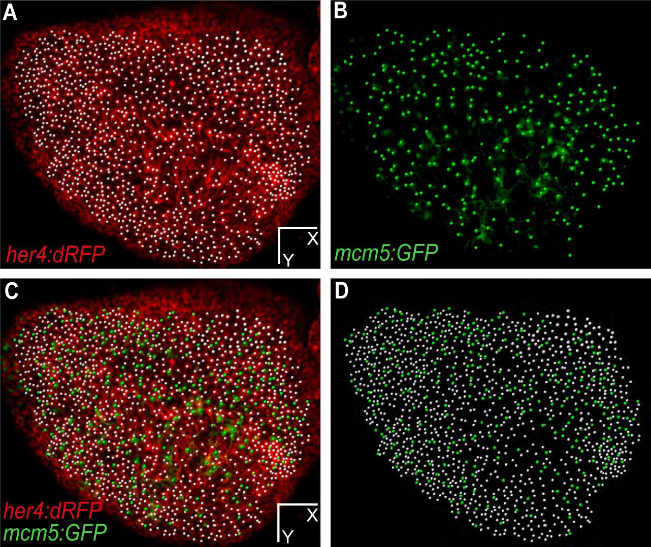
Fig. S10 Plotting of adult neural stem cells and proliferating progenitors at time point 0 on a live image. Dorsal view of one hemisphere of a her4:dRFP;mcm5:eGFP;casper transgenic fish (individual fish named piwi) imaged at t0. (A) red channel only (RG cells) with corresponding plots (white dots), (B) green channel only (proliferating cells), (C) merged red and green fluorescence view with corresponding plots for RG (white) and proliferating progenitors (green), (D) plots only (as in Fig. 2E).
Scale bars: 80 µm (A-D).
Acknowledgments
This image is the copyrighted work of the attributed author or publisher, and
ZFIN has permission only to display this image to its users.
Additional permissions should be obtained from the applicable author or publisher of the image.
Full text @ Development