
|
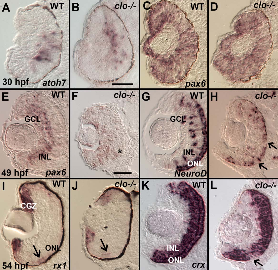
Fig. 8
Irregular expression of specific retinal transcription factors in cloche mutant embryos. A–D: At 30 hpf atoh7/ath5 is expressed in a ventral cluster of retinal progenitor cells in both wild-type (A) and clo-/- (B), and pax6a is expressed in all retinal progenitors in both wild-type (C) and clo-/- (D) embryos. E,F: At 49 hpf pax6a is expressed in the ganglion cell layer (GCL) and inner half of the inner nuclear layer (INL) of wild-type embryos (E), but is more diffusely distributed (such as in the cells near the asterisk) and weakly expressed in clo-/- (F). G,H: At 49 hpf, neurod1 is expressed in the ONL, and in radial clusters of cells in the INL and occasionally the GCL in wild-type (G), but is reduced in distribution in all of these locations in clo-/- (H), resulting in patches of ONL lacking neurod1 (arrows). I,J: At 54 hpf, rx1 is expressed in the circumferential germinal zone (CGZ) and weakly in the emerging outer nuclear layer (ONL, arrows) in WT (I) and clo-/- (J); K,L: At 54 hpf crx is expressed in the ONL and the outer half of the INL in wild-type (K), and shows a similar distribution and hybridization intensity in clo-/- (L), but with occasional patches of ONL lacking crx expression (arrow). Scale bars = 50 µm in B (applies to A–D(; F (applies to E–L). Sections in panels E–H, and K–L were derived from embryos treated with PTU and so do not have melanin pigment within the retinal pigmented epithelium.