
|
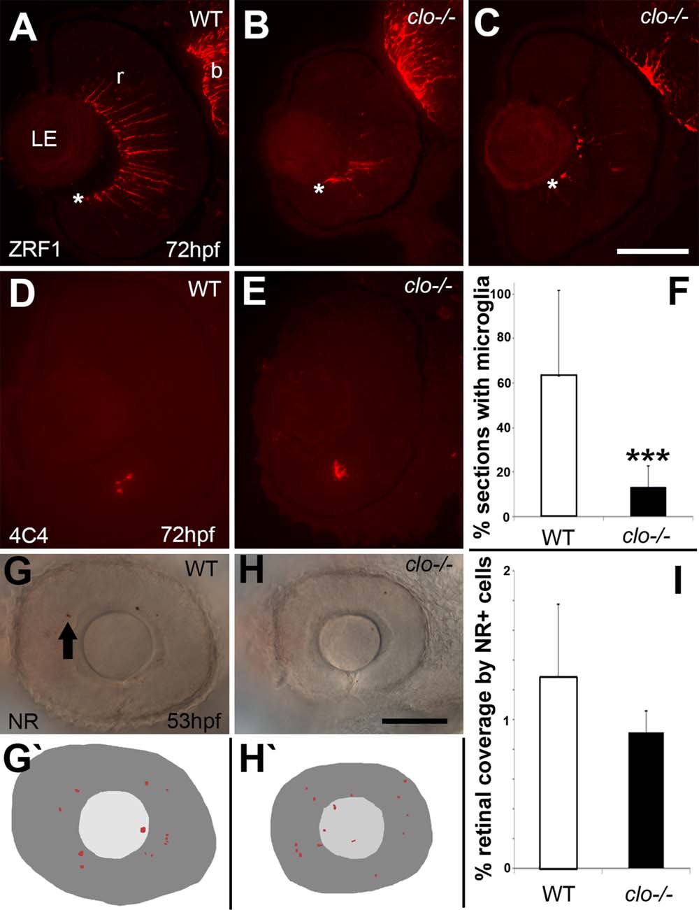
Fig. 7
Abnormal differentiation of Müller glia, and reduced microglia in cloche mutant embryos. A–C: Immunofluorescence images of 72 hpf retinas of wild-type (A) and clo-/- (B,C) embryos stained with the ZRF1 antibody detecting GFAP, specific to Müller glia. * indicates vitreal surface of retina (r), where Müller glia establish endfeet. b, brain. D,E: Immunofluorescence images of 72 hpf retinas of wild-type (D) and clo-/- (E) embryos stained with the 4C4 antibody that labels microglia. F: The % of cryosections per eye that contained microglia was significantly reduced in clo-/- embryos as compared with wild-type embryos (***P < 0.0001). G,H: Neutral red uptake in retinal cells of wild-type (G) and clo-/- (H) embryos from 49 to 53 hpf. G′–H′: Tracings of flattened projections of neutral red (NR) stained wild-type (G′) and clo-/- (H′) eyes; arrow in G shows an NR+ cell. I: Percent retinal coverage by neutral red+ profiles. LE, lens. Scale bar = 50 µm in C (applies to A–E); 50 µm in H (applies to G,H′).