
|
Fig. 3
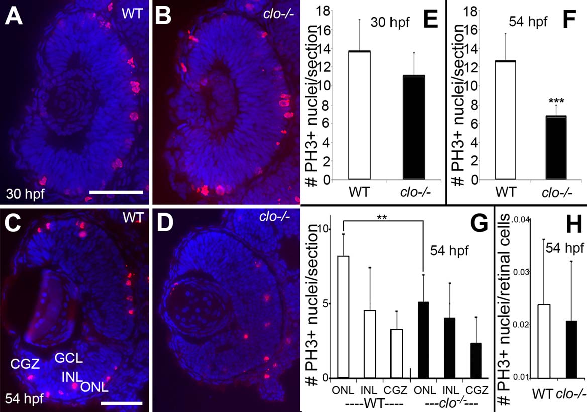
Retinal progenitor proliferation in wild-type and clo mutant embryos. A–D: Immunofluorescence images of wild type (A,C) and clo/ (B,D) retinas stained with anti-phosphohistone 3 (PH3) and counterstained with DAPI (blue); samples obtained at 30 hpf (A,B) and 54 hpf (C,D). E–G: Numbers of PH3+ nuclei/section are not significantly different in clo-/- vs. wild-type retinas at 30 hpf (E), but are significantly different at 54 hpf (F; P < 0.001); these differences are most evident in the outer nuclear layer (G; ONL; P < 0.001), but not in the inner nuclear layer (INL) or circumferential germinal zone (CGZ). H: Mitotic index (# PH3+ nuclei/total number of retinal cells per section) is not significantly different in clo-/- as compared to wild-type. Scale bars = 50 µm in A (applies to A,B); C (applies to C,D).