
Image
|
Figure Caption
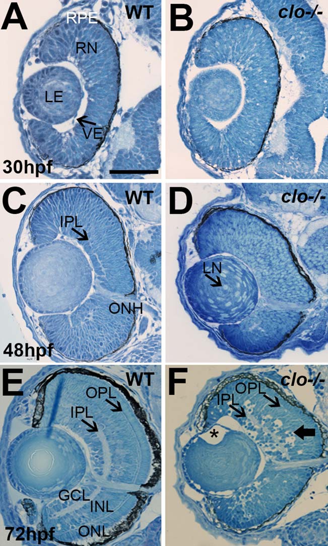
Fig. 2
A–F: Histology of wild-type (A,C,E) and clo-/- (B,D,F) eyes at 30 hpf, 48 hpf, and 72 hpf. RPE, retinal pigmented epithelium; RN, retinal neuroepithelium; LE, lens; VE, vascular endothelial cells; IPL, inner plexiform layer; ONH, optic nerve head; GCL, ganglion cell layer; INL, inner nuclear layer; ONL, outer nuclear layer; OPL, outer plexiform layer. In F, asterisk (*) denotes gap between lens epithelium and developing lens fibers; wide arrow points to pyknotic cells in INL. Scale bar = 50 µm in A (applies to all).
Acknowledgments
This image is the copyrighted work of the attributed author or publisher, and
ZFIN has permission only to display this image to its users.
Additional permissions should be obtained from the applicable author or publisher of the image.
Full text @ Dev. Dyn.