
Image
|
Figure Caption
Fig. 6
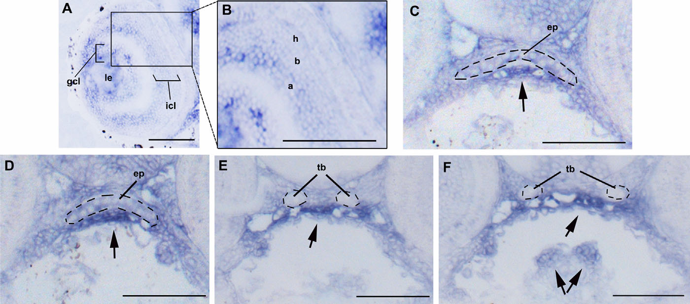
Spatial localization of rxfp2l transcript outside the zebrafish brain. A: Detail of transverse section showing the eye of zebrafish larvae (72 hpf). B: Magnification of inner cell layer as indicated in A. C–F: Detail of transverse sections showing the pharyngeal arches region of zebrafish larvae (72 hpf). Black arrow indicates rxfp2l-expressing cells in the epithelium. Scale bar: 50 µm. a, amacrine cells; b, bipolar cells; Ep, ethmoid plate; gcl, ganglion cell layer; h, horizontal cells; icl, inner cell layer; le, lens; tb, trabeculae.
Figure Data
Acknowledgments
This image is the copyrighted work of the attributed author or publisher, and
ZFIN has permission only to display this image to its users.
Additional permissions should be obtained from the applicable author or publisher of the image.
Full text @ J. Exp. Zool. B Mol. Dev. Evol.