
|
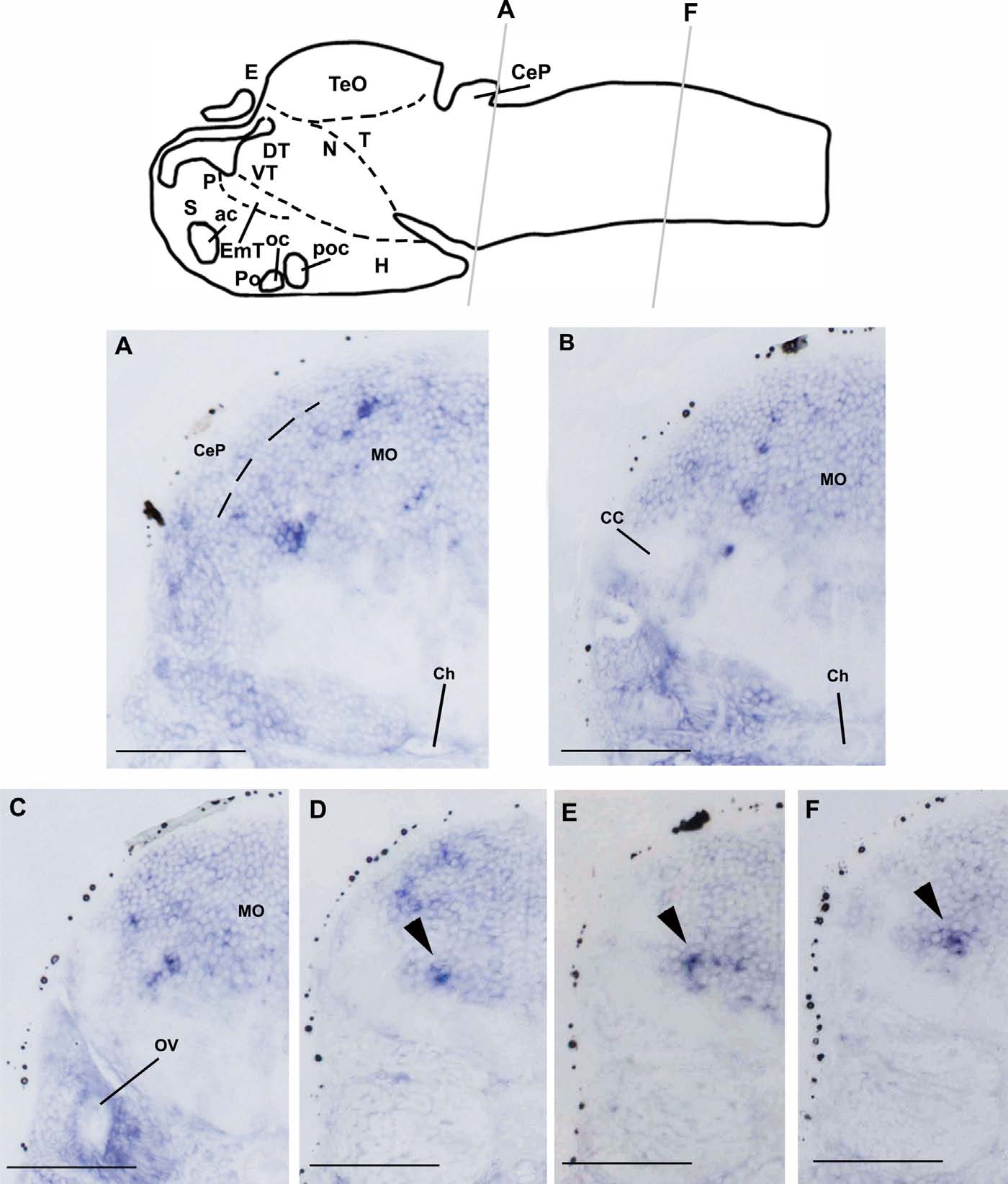
Fig. 5
Spatial localization of rxfp2l transcript in the caudal region of zebrafish larval brain. A–F: Transverse sections of hybridized zebrafish larvae (72 hpf) as indicated in the schematic drawing of the zebrafish larval brain. Black arrowhead indicates restricted rxfp2l-expressing cell cluster in the medulla oblongata. Scale bar: 50 µm. ac, anterior commissure; CC, cerebellar crest; CeP, cerebellar plate; Ch, chorda dorsalis; DT, dorsal thalamus; E, epiphysis; EmT, eminentia thalami; H, hypothalamus; MO, medulla oblongata; N, region of the nucleus of medial longitudinal fascicle; oc, optic chiasma; OV, otic vesicle; P, pallium; Po, preoptic region; poc, postoptic region; S, subpallium; T, midbrain tegmentum; TeO, optic tectum; VT, ventral thalamus.