
|
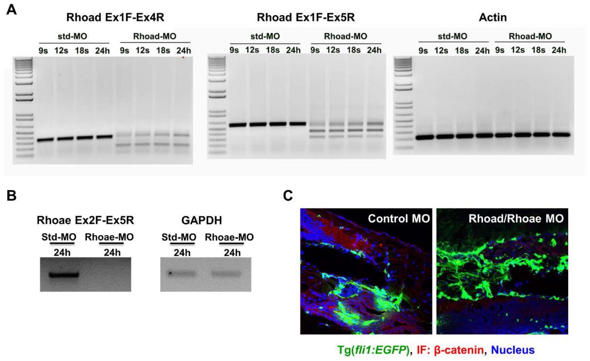
Fig. S4 MO-mediated RhoC ablation decreases β-catenin in the vessels of zebrafish. (A-B) RhoC (Rhoad/Rhoae) or control (std) MOs were injected into 1-2 cell stage Tg(Fli:GFP) zebrafish embryos. Embryos were collected at the indicated times, RNA was collected using an RNeasy Plus Mini Kit (Qiagen) and converted to cDNA, PCR was performed using the indicated oligonucleotide pairs, and visualized after running on an agarose gel. (A) Oligonucleotides were used to amplify Rhoad Exon 1-4 (A, left), Rhoad Exon 1-5 (A, middle) and loading control β-actin (A, right). (B) Oligonucleotides were used to amplify Rhoae Exon 2-5 (B, left) and loading control GAPDH (B, right). (C) RhoC (Rhoad/Rhoae) or control MOs were injected into 1-2 cell stage Tg(Fli:GFP) zebrafish embryos. At 3 dpf, the zebrafish were formalin-fixed, placed in OCT, frozen, sectioned with a microtome onto slide, stained with a primary β-catenin antibody followed by a corresponding secondary Alexa-Fluor 568 (red) antibody, and visualized using a Zeiss LSM 780 confocal microscope. Green: Tg(Fli:GFP); Red: β-catenin; Blue: DAPI-stained nuclei.