
Fig. 2
- ID
- ZDB-IMAGE-151110-2
- Antibodies
- Publication
- Kimmel et al., 2015 - Diabetic pdx1-mutant zebrafish show conserved responses to nutrient overload and anti-glycemic treatment
- All Figures
- Figures for Kimmel et al., 2015
|
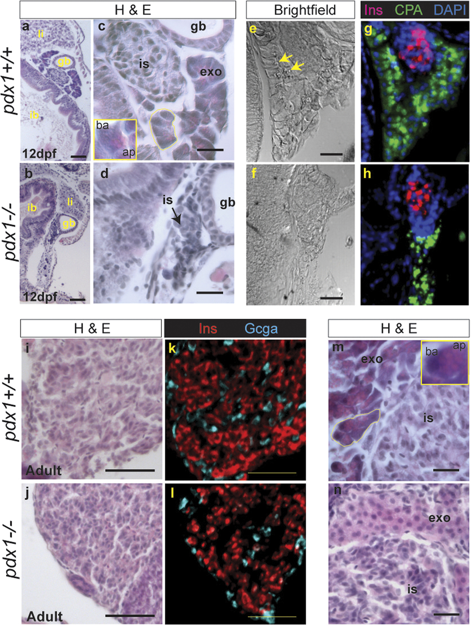
Fig. 2 Pancreatic histology of pdx1 mutants.
(a–d) Gut region morphology in longitudinal-oblique sections of 12 dpf wild type (a,c), and pdx1 mutant (b,d) larvae stained with hemotoxylin and eosin (H&E). (a–d) Pancreatic tissue is located between the intestinal bulb (ib) and gall bladder (gb). (li, liver) (c) In wild type embryos, exocrine cells (exo) adjacent to the islet (is) show clear polarity and acinar organization (yellow outline). Inset, close-up of single acinar cell, showing basophilic basal region (ba) and eosinophilic apical region (ap), where secretory granules are located. (d) In pdx1 mutants cell polarity and acini are not apparent. In brightfield images of an adjacent region, exocrine secretory granules can be discerned in the wild type pancreas (e, arrows) in the region immunostained for CPA (green) (g). (f,h) CPA-positive exocrine tissue in the pdx1 mutant appears disorganised. Beta cells of the islet are labeled by immunostaining for Ins (red). Nuclei are counterstained with DAPI (blue). ((a,b), scale bar = 50µm; (c–f), scale bar = 20µm) In adults, Ins + cells (red) occupy the core of the islet, while Gcga+ cells (cyan) are found mostly at the periphery in both controls and pdx1 mutants (i–l). Scale bar=50µm. (m) Polarized exocrine cells (exo) arranged in acini (yellow outline) are found adjacent to the islet in controls Inset, close-up of a single acinar cell, showing basally (ba) located nucleus and eosinophilic apical region (ap), containing secretory granules. (n) In pdx1 mutants, exocrine cells appear unpolarized and lack acinar organization. ((m,n) scale bar = 20µm)