
Fig. 4
- ID
- ZDB-IMAGE-151105-7
- Publication
- Cortés-Campos et al., 2015 - Zebrafish adult-derived hypothalamic neurospheres generate gonadotropin-releasing hormone (GnRH) neurons
- All Figures
- Figures for Cortés-Campos et al., 2015
|
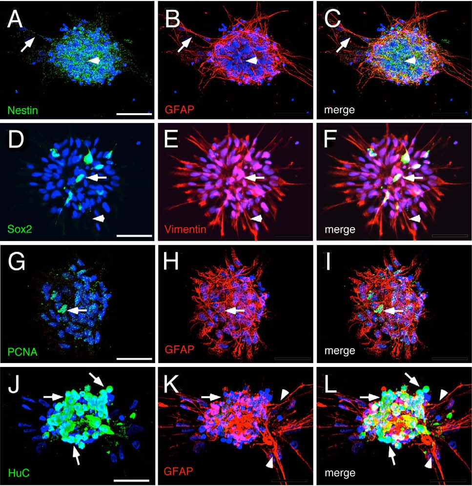
Fig. 4
Hypothalamic progenitors generate glial and neuronal cells. (A-F) Neurospheres differentiated after 7days in vitro. (A-C) Differentiated cells surround the core of cells positive for nestin (arrow head) and extend cellular projections that are reactive to GFAP (arrows). (D-F) The reduced number of Sox2 positive cells (arrows) and the high number of cell processes reactive to Vimentin (arrow heads) suggests that progenitors have differentiated into glial cells. (G-L) Neurospheres reactive to GFAP show (G-I) a reduced number of cells positive for PCNA (arrows), (J-L) high number of neurons (arrows) and differentiated glial cells (arrow heads). Scale bar 25µm, n=3 plates derived from different cell cultures.