
Fig. S1
- ID
- ZDB-IMAGE-151030-6
- Publication
- Antonio et al., 2015 - The wound inflammatory response exacerbates growth of pre-neoplastic cells and progression to cancer
- All Figures
- Figures for Antonio et al., 2015
|
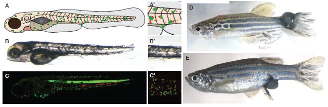
Fig. S1
Standard larval model for studying immune cell:pre-neoplastic cell interactions.
A. Schematic to illustrate the larval model, whereby oncogenic Human RasG12V is used to drive cell transformation in a tissue specific manner, in melanocytes and mucus-secreting goblet cells by a kit-a enhancer trap (ET30).
B. DIC microscopy of a 5dpf kita:RasG12VeGFP zebrafish larvae.
C. Confocal laser scanning microscopy image of a 5dpf kita:RasG12VeGFP; LysC:dsRed zebrafish larvae. (A′, B′, C′) Zoom in of the area around the cloaca to illustrate the region frequently imaged.
D. and E Adult fish occasionally develop large pigmented tumours, particularly at exposed sites such as the ventral fin by the cloaca or tail.