
IMAGE
Fig. S4
- ID
- ZDB-IMAGE-151029-25
- Publication
- Chen et al., 2015 - Transient laminin beta 1a Induction Defines the Wound Epidermis during Zebrafish Fin Regeneration
- All Figures
- Figures for Chen et al., 2015
Image
|
Figure Caption
Fig. S4
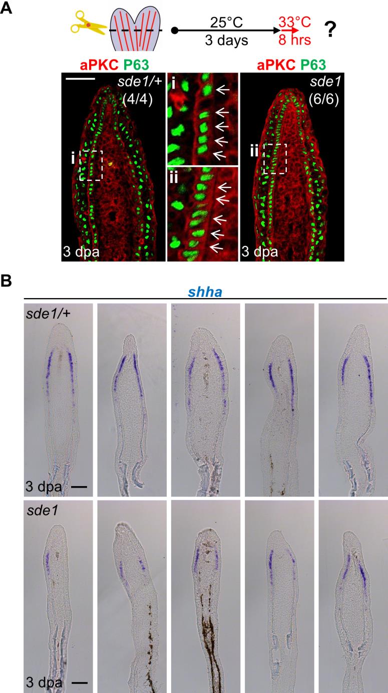
Eight hours of 33°C treatment is sufficient to alter aPKC localization and reduce shha expression in sde1 regenerates.
(A) Antibody co-staining for aPKC (red) and P63 (green) in longitudinal sections of sde1/+ and sde1 fin regenerates after 8 hours of 33°C treatment at 3 dpa, indicating rapid loss of basal cell polarity. Scale bars, 50 µm. (B) shha RNA expression is also reduced in sde1 regenerates after 8 hours of 33°C treatment (n = 5 each). Scale bars, 100 µm.
Acknowledgments
This image is the copyrighted work of the attributed author or publisher, and
ZFIN has permission only to display this image to its users.
Additional permissions should be obtained from the applicable author or publisher of the image.
Full text @ PLoS Genet.