
Fig. 9
- ID
- ZDB-IMAGE-151026-7
- Publication
- Mitchell et al., 2015 - Retinoic Acid Signaling Regulates Differential Expression of the Tandemly-Duplicated Long Wavelength-Sensitive Cone Opsin Genes in Zebrafish
- All Figures
- Figures for Mitchell et al., 2015
|
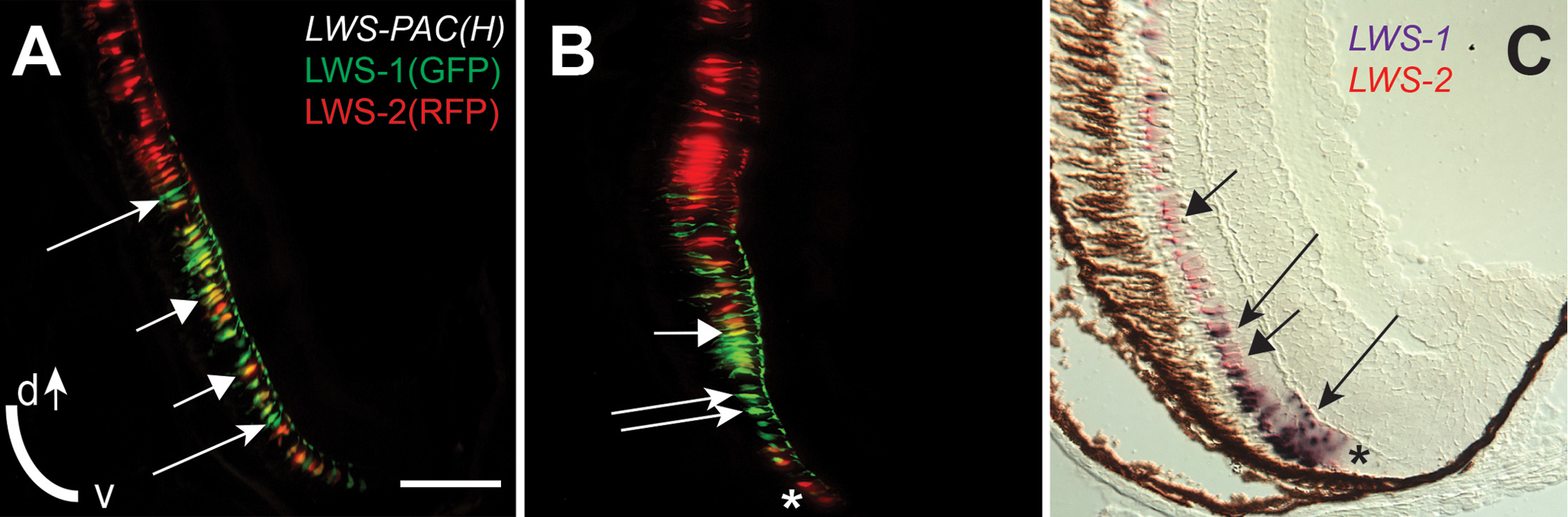
Fig. 9
An “LWS Transition Zone” exists in ventral retina of juvenile fish.
Sections of retina from one-month old juvenile retinas are shown. A and B. Two different sections from separate LWS:PAC(H) transgenic fish showing LWS1:GFP+ cones (green fluorescence; long arrows), LWS2:RFP+ cones (red fluorescence), and cones co-expressing LWS1:GFP and LWS2:RFP (yellow, short arrows). B. The asterisk (*) indicates a cone expressing only LWS2:RFP at the ventral periphery. C. Dual in situ hybridization using cDNA probes for LWS1 (purple) and LWS2 (red/pink). Cones expressing LWS1 only are indicated by long arrows; cones co-labeled with both probes are indicted by short arrows. The asterisk (*) indicates an LWS2 singly labelled cone at the ventral periphery. Scale bar in A (applies to all) = 25 µm; v, ventral; d, dorsal.