Image
Figure Caption
Fig. S2
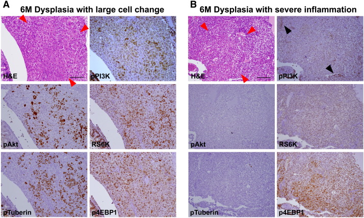
Activation of PI3K pathway in serial sections (same sections with Supplementary Figure 1). (A) Dysplasia with large cell changes showing expression of active components of PI3K pathway. (B) Dysplasia with robust inflammation. Contrary to (A), pPI3K expression is noted in inflammatory cells (black arrowheads) with faint patch expression in hepatocytes. Downstream components of PI3K pathway are not activated, showing nil pAkt, RS6k, or pTuberin expression but p4EBP1 expression.
Acknowledgments
This image is the copyrighted work of the attributed author or publisher, and
ZFIN has permission only to display this image to its users.
Additional permissions should be obtained from the applicable author or publisher of the image.
Full text @ Neoplasia

