Fig. 2
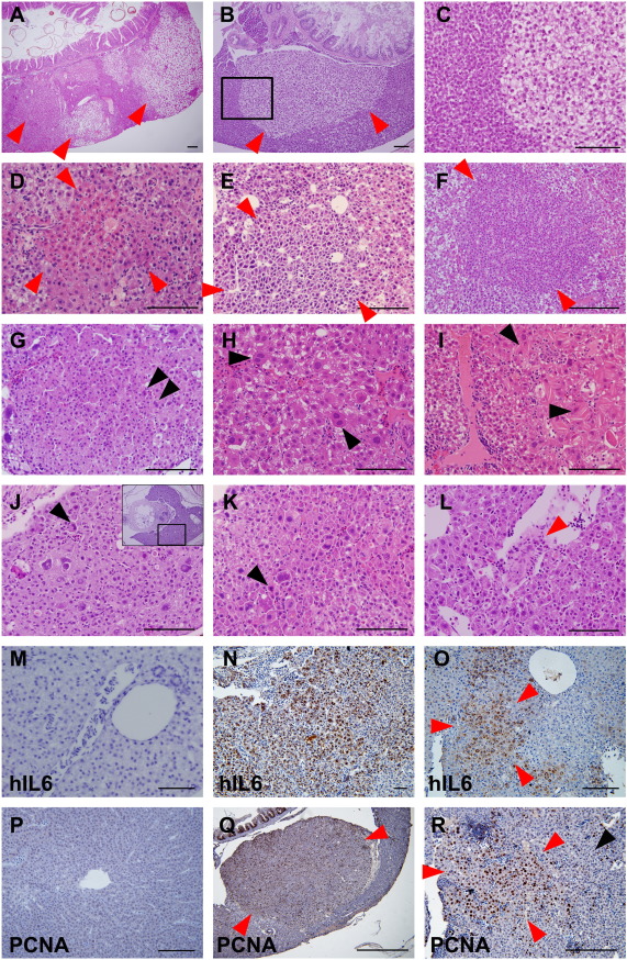
H&E staining of liver tumor progression in the transgenic zebrafish. (A–C) Two-month-old zebrafish. (D–I, M–R) Three-month-old zebrafish. (J–L) Six-month-old zebrafish. (M) and (P) are control liver images of Tg(LFABP:Gal4VP16; UAS:RFP), and others are from Tg(LFABP:Gal4VP16;UAS:RFP;UAS:hIL6,Cmcl2:GFP). All red arrowheads indicate dysplastic foci. (A) Multiple foci of hepatocellular alteration (red arrowheads). Clear cell change was most frequently observed. (B) Clear cell focus with scanty inflammation in the surrounding liver. (C) A high-power observation of the boxed area of (B) showing nuclear pleomorphism. (D) Eosinophilic focus of hepatocellular alteration. (E) Basophilic cell change showing cytological atypia with nuclear pleomorphism and high nucleocytoplasmic ratio. (F) Small cell change with increased cell density and high nucleocytoplasmic ratio. (G) Small cell change showing nuclear pleomorphism and occasional multinucleation (black arrowheads). (H) Large cell change with nuclear pleomorphism and frequent multinucleated cells (black arrowheads). (I) Dysplastic changes with eosinophilic mutilamellar structure (black arrowhead). (J–L) Overt HCCs showing marked nuclear and cytological pleomorphism of tumor cells at 6 (J) and 9 (K and L) months. (J) High-magnification view of boxed area of inset. Occasional mitoses are seen (black arrowheads). Occasionally, tumor cells invaded intrahepatic vessels, forming tumor thrombi (red arrowhead in L). (M–O) IHC against hIL6. Contrary to control liver (M), majority of the hepatocytes in M were found to highly express hIL6. (O) Hepatocytes in a dysplastic focus (red arrowhead) showing a robust expression of hIL6. (P–R) IHC against PCNA. (P) Control. (Q) PCNA expression was highly positive in the cells in the dysplastic nodule which appears as bulging mass (red arrowheads). (R) The hepatocytes at dysplastic focus (red arrowheads) showed more frequent positivity to PCNA expression than the ones at nondysplastic area (black arrowhead). Bars, 50 µm.

