
IMAGE
Fig. 3
- ID
- ZDB-IMAGE-151026-1
- Genes
- Antibodies
- Publication
- Horng et al., 2015 - Aquaporin 1 Is Involved in Acid Secretion by Ionocytes of Zebrafish Embryos through Facilitating CO2 Transport
- All Figures
- Figures for Horng et al., 2015
Image
|
Figure Caption
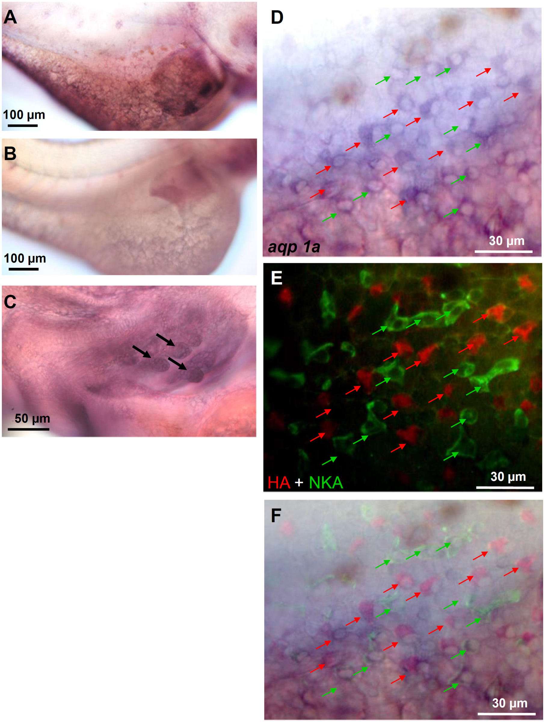
Fig. 3
Localization of aqp1a.1
Triple-labeling of aqp1a.1 mRNA (with in situ hybridization), H+-ATPase (HA), and Na+/K+-ATPase (NKA) (with immunofluorescence) in 3-dpf embryos. The aqp1a.1 antisense probe labeled ionocytes in the yolk-sac skin of embryos (A, D). Those signals were not found in the negative control with an aqp1a.1 sense probe (B). aqp1a.1 signals (arrows) were also found in developing gills (C). The yolk-sac aqp1a.1 signals (D) were co-localized with HA (red arrows in E) and NKA (green arrows in E). A merged image of (D) and (E) is shown in (F).
Figure Data
Acknowledgments
This image is the copyrighted work of the attributed author or publisher, and
ZFIN has permission only to display this image to its users.
Additional permissions should be obtained from the applicable author or publisher of the image.
Full text @ PLoS One