Fig. 8
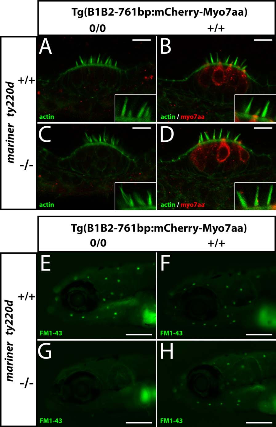
Compensation of mariner ty220d / mutation by expression of mCherry-Myo7aa fusion protein under the control of B1B2mB-761bp DNA element leads to restoration of sensory hair cell function. (A–D) Recovery of normal hair bundle morphology was observed by staining with Alexa 488-Phalloidin (green) and detection of mCherry-Myo7aa fusion protein expressed in Tg(B1B2mB-761bp:mCherry-Myo7aa) transgene was detected by immunofluorescence (red) in 5 dpf embryos. (A,C) embryos do not express Tg(B1B2mB-761bp:mCherry-Myo7aa) transgene whereas embryos depicted in (B,D) do express the fusion protein. (A and B) embryos carrying endogenous mar ty220d +/+ genotype; (C and D) embryos carrying endogenous mar ty220d -/- genotype. Inlays are high magnification views of a few hair bundles showing the typical shape observed within the imaged lateral cristae. Fixed embryos mounted in agarose were observed on a SP5 Leica confocal microscope. Images are single z-axis sections. Scale bar, 10 µm, applies for the lower magnification of the whole cristae. (E–H) Recovery of FM 1-43 uptake in lateral line hair cells of mar ty220d -/- complemented with mCherry–Myo7aa fusion protein expressed under the control of B1B2mB761bp DNA fragment. (E, G) embryos do not express Tg(B1B2mB-761bp:mCherry-Myo7aa) transgene whereas embryos depicted in (F,H) do express the fusion protein. (E and F) embryos carrying endogenous mar ty220d +/+ genotype; (G and H) embryos carrying endogenous mar ty220d -/- genotype. 5 dpf live embryos were observed on an Olympus Macro Zoom. Scale bar = 250 µm.

