
|
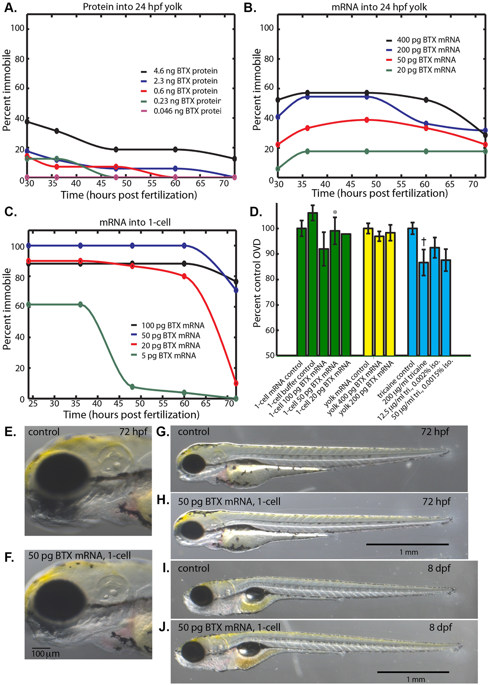
Fig. 2
α-bungarotoxin immobilizes embryos while permitting normal development.
(A) Percent of embryos immobile after injection of α-bungarotoxin protein (0.046–4.6ng) into the yolk at 24 hpf. (B) Percent of embryos immobile after injection of α-bungarotoxin mRNA (20–400 pg) into the yolk at 24 hpf. (C) Percent of embryos immobile after injection of of α-bungarotoxin mRNA (5–100 pg) into the 1-cell zygote. (D) Percent control OVD at 72 hpf for injection of α-bungarotoxin mRNA into the 1-cell zygote (green), into the yolk (yellow), and reference anesthetic treatments that permitted long-term immobilization (blue). (*) Not significantly different from control, Mann-Whitney-Wilcoxon two tailed P-value 0.87. (†) Significantly different from control, Mann-Whitney-Wilcoxon two tailed P-value 0.0011. (E, G) Control embryo at 72 hpf that was injected with 50 pg of membrane-citrine mRNA into the 1-cell zygote. (F, H) 72 hpf embryo that was injected with 50 pg of α-bungarotoxin mRNA into the 1-cell zygote. (I) Control larva at 8 days post fertilization (dpf) injected with 50 pg of membrane-citrine mRNA into the 1-cell zygote. (J) 8 dpf larva that was injected with 50 pg of α-bungarotoxin mRNA into the 1-cell zygote.