
Fig. S11
- ID
- ZDB-IMAGE-151009-31
- Publication
- Jia et al., 2015 - Mutation of kri1l causes definitive hematopoiesis failure via PERK-dependent excessive autophagy induction
- All Figures
- Figures for Jia et al., 2015
|
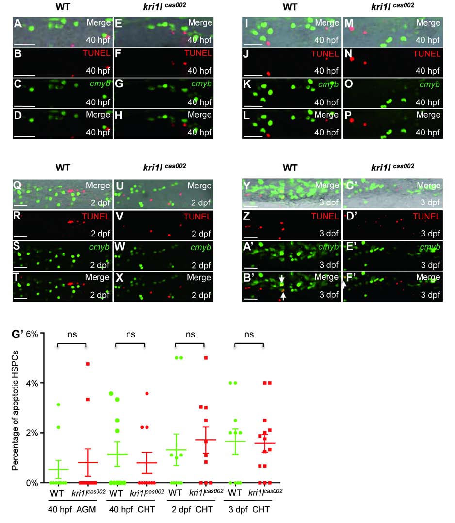
Fig. S11 Percentage of apoptotic HSPC in kri1lcas002 mutants and WT siblings. Representative confocal images of kri1lcas002 mutants and WT siblings after TUNEL assays co-staining with EGFP antibody (in Tg(cmyb:egfp) background) in different time points and regions, including the AGM at 40 hpf (A-H) (A and E are bright-field images overlaid with fluorescent staining. Scale bars, 25 µm.); the CHT at 40 hpf (I-P) (I and M are bright-field images overlaid with fluorescent staining. Scale bars, 25 µm.), at 2 dpf (Q-X) (Q and U are bright-field images overlaid with fluorescent staining. Scale bars, 25 µm.), and at 3 dpf (Y-F′) (Y and C′ are bright-field images overlaid with fluorescent staining. Scale bars, 25 µm.). (G′) Statistic analysis of the apoptotic HSPC percentage (the proportion of apoptotic HSPC in total HSPCs) in kri1lcas002 mutants and wild-type siblings during hematopoietic development (40 hpf-3 dpf). Ne10 for each group. 40 hpf AGM, p=0.6824; 40 hpf CHT, p=0.5971; 2 dpf CHT, p=0.6433; 3dpf CHT, p=0.9122. Error bars represent standard error of the mean (SEM). **, P≤0.01; ***, P≤0.001 (Student t test).