
|
Fig. S3
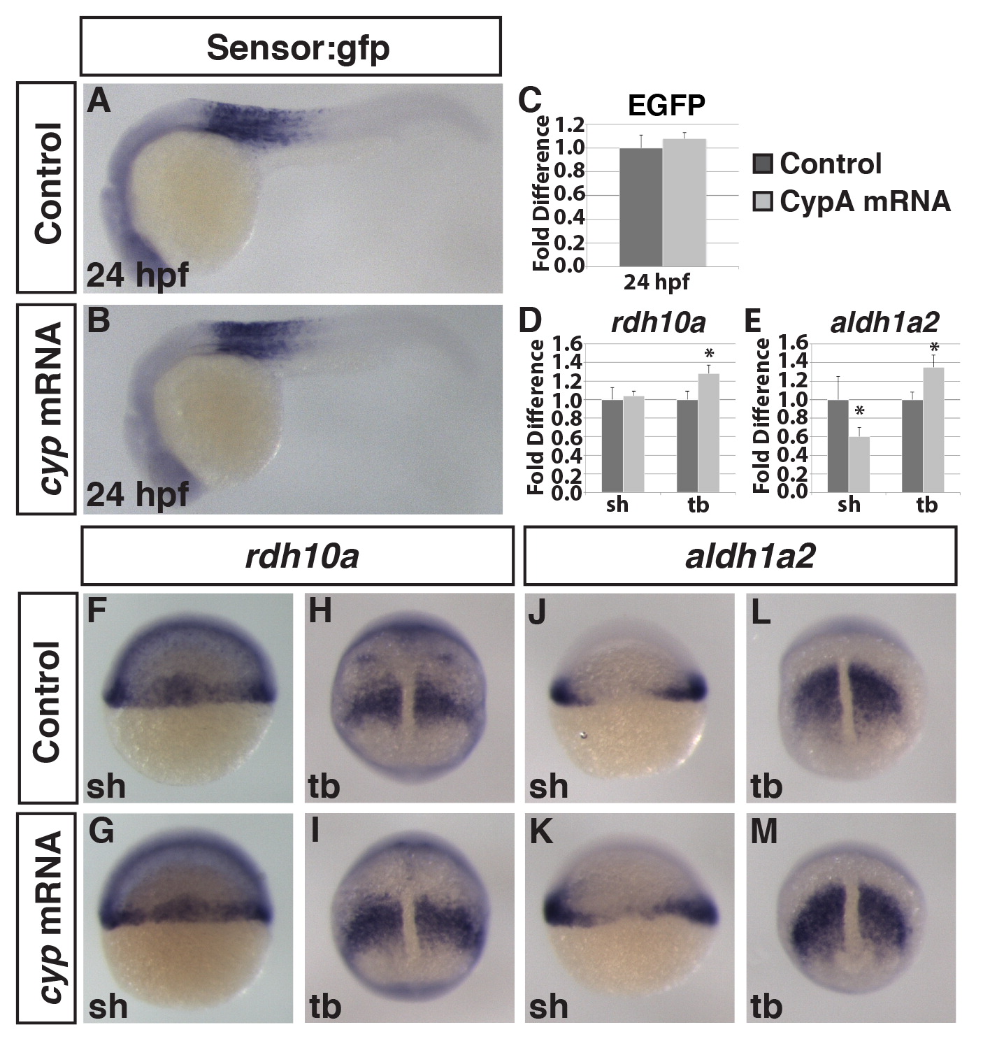
Injection of cyp26a1 mRNA is not sufficient to induce a dramatic loss of RA signaling. (A,B) ISH of GFP expression in Tg(β-actin:GDBD-RLBD);(UAS:EGFP) embryos injected with cyp26a1 (cyp) mRNA show no difference in EGFP. (C) qPCR for EGFP in Tg(β-actin:GDBD-RLBD);(UAS:EGFP) embryos show no difference in EGFP expression between Cyp26a1 mRNA compared to WT siblings. (D,E) Quantification of (F–M) by RT-qPCR shows that both rdh10a and aldh1a2 are increased by tailbud. (F,G,H,I) ISH for rdh10a shows embryos injected with cyp26a1 mRNA have no difference at shield stage but are increased by tailbud. (J,K,L,M) ISH for aldh1a2 shows cyp mRNA injection has no effect at shield stage but leads to an increase in aldh1a2 by tailbud. Asterisks indicate statistically different from controls (p<0.05) using a Student′s t-test.
Reprinted from Developmental Biology, 405(1), Rydeen, A., Voisin, N., D'Aniello, E., Ravisankar, P., Devignes, C.S., Waxman, J.S., Excessive feedback of Cyp26a1 promotes cell non-autonomous loss of retinoic acid signaling, 47-55, Copyright (2015) with permission from Elsevier. Full text @ Dev. Biol.