
|
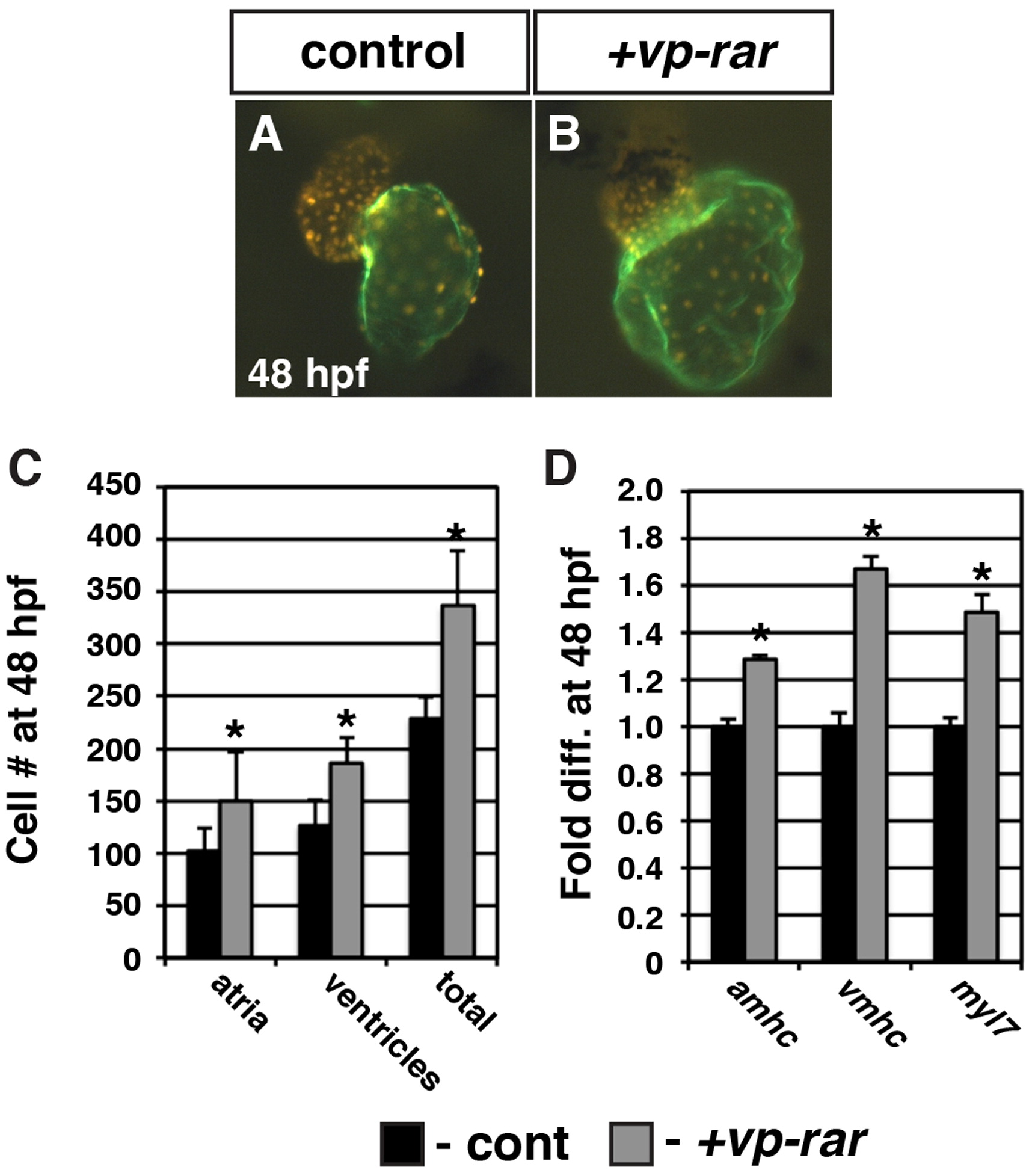
Fig. 2
VP-RAR expression promotes increased cardiomyocyte number. (A, B) The hearts from vp-rar mRNA injected Tg(myl7:DsRed-NLS) embryos are enlarged relative to siblings. Images are frontal views. Red alone indicates ventricular cells. Atrium is indicated in green (S46 antibody). (C) Cell counts of vp-rar mRNA injected embryos at 48 hpf reveal an increase in both atrial and ventricular cardiomyocytes relative to control sibling embryos. Control n=10. Vp-rar injected n=7. (D) RT-qPCR at 48 hpf of cardiac markers amhc, vmhc, and myl7 shows increased expression in vp-rar mRNA injected embryos compared to control siblings. Asterisks indicate statistically different from control (p<0.05) using Student′s t-test.
Reprinted from Developmental Biology, 405(1), Rydeen, A., Voisin, N., D'Aniello, E., Ravisankar, P., Devignes, C.S., Waxman, J.S., Excessive feedback of Cyp26a1 promotes cell non-autonomous loss of retinoic acid signaling, 47-55, Copyright (2015) with permission from Elsevier. Full text @ Dev. Biol.