
|
Fig. 6
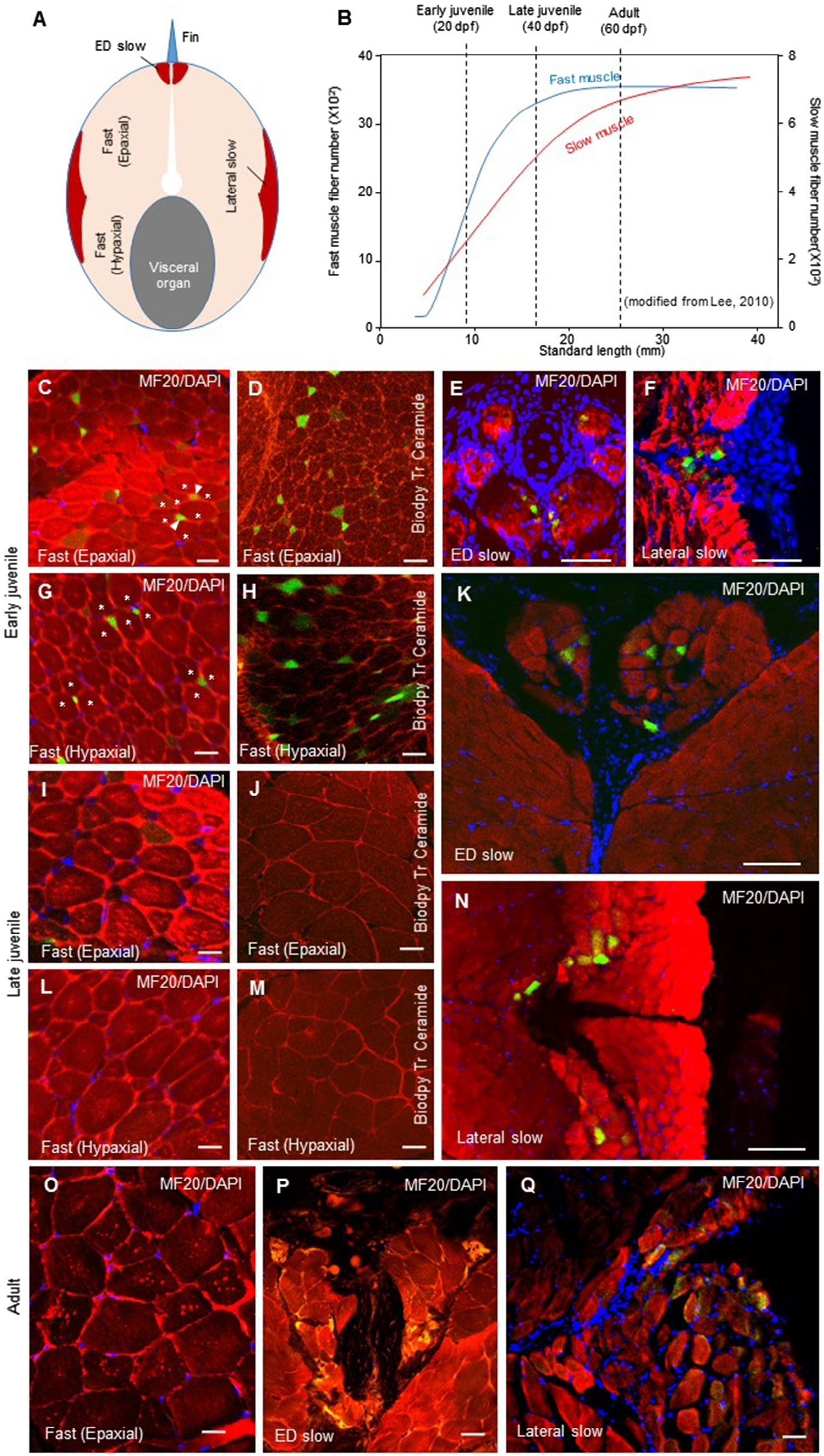
Localization of EGFP-expressing myotomal muscle fibers in Tg:MYHM2528-1:EGFP at early juvenile (20 dpf, 8 mm standard length (SL), late juvenile (40 dpf, 17 mm SL) and adult (60 dpf, 25 mm SL) stages). (A) Schematic of a cross-section of the myotomal region. Slow muscle is distributed throughout the base of the dorsal fin (erector and depressor (ED) slow) and lateral surface (lateral slow). (B) Body size-related increase in muscle fiber numbers in slow and fast muscles of zebrafish. Data cited from Lee (2010). (C–H) Cross-sectional view of the myotomal region at the early juvenile stage. Muscle fibers were stained with MF20 (C,E,F,G) and BIODIPY TR Ceramide (D,H). EGFP expression was observed in small diameter fast muscle fibers (C,D,G,H), the inner part (near the septum between slow and fast muscles) of lateral slow (E) and ED slow (F) muscles. In panel C and G, an EGFP-positive small ‘neonatal’ muscle fiber and the surrounding large ‘old’ fibers are marked by an arrowhead and asterisks, respectively. (I–N) Cross-sectional view of the myotomal region at the late juvenile stage. Muscle fibers were stained with MF20 (I,K,L,N) and BIODIPY TR Ceramide (J,M). EGFP expression was not observed in fast muscle (I,G,L,M). In the inner portions of lateral slow (K) and ED slow (N) muscle fibers expressed EGFP at the early juvenile stage. (O–Q) Cross-sectional view of the myotomal region at the adult stage. Muscle fibers were stained with MF20. In consist with late juvenile stage, EGFP expression was not observed in fast muscle (O) but expressed at the inner portions of ED slow (P) and lateral slow (Q) and muscle fibers. Arrow heads indicate positive EGFP expression. Scale bar: 50 µm.
Reprinted from Mechanisms of Development, 137, Ahammad, A.K., Asaduzzaman, M., Asakawa, S., Watabe, S., Kinoshita, S., Regulation of gene expression mediating indeterminate muscle growth in teleosts, 53-65, Copyright (2015) with permission from Elsevier. Full text @ Mech. Dev.