
Fig. 4
- ID
- ZDB-IMAGE-151005-19
- Genes
- Publication
- Mendelson et al., 2015 - Maternal or zygotic sphingosine kinase is required to regulate zebrafish cardiogenesis
- All Figures
- Figures for Mendelson et al., 2015
|
Fig. 4
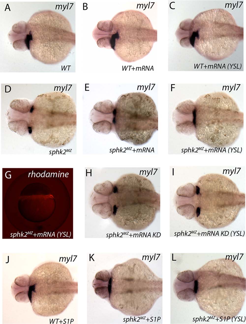
Expression of Sphk2 in YSL can rescue normal cardiogenesis. Shown are representative embryos following in situ hybridization for myl7 transcripts at 32 hpf. (A) Wildtype (WT) embryos (25/25, 100%) display normal cardiac development. (B) Injection of 250 pg of sphk2 mRNA into the single cell (65/65, 100%) or (C) targeted to the YSL (51/51, 100%) does not alter cardiac development. (D) All maternal/zygotic sphk2MZ embryos show the cardia bifid phenotype (0/40 embryos are normal, 0%). (E) Injection of 250 pg sphk2 mRNA into the 1-cell stage embryos was able to rescue the bifid phenotype as shown by the presence of cardiac tissue at the midline (7/44 embryos, 16%). (F) Targeting 250 pg sphk2 mRNA specifically into the YSL (confirmed by co-injection of rhodamine dextran tracer, as documented in G) was able to rescue the bifid phenotype (19/88 embryos, 22%). (H) In contrast, injection of mRNA encoding a predicted kinase dead Sphk2 (KD) into the fertilized egg (56/56, 100%) or (I) targeted to the YSL (43/43, 100%) failed to rescue the bifid phenotype. (J) Injection of 1 ng S1P in the fertilized egg (47/47, 100%) or YSL (47/47, 100%, not shown) did not alter cardiac development of wildtype embryos, whereas (K) injection of 1 ng S1P into the fertilized egg (15/51, 29%) or (L) targeted to the YSL (9/32, 28%) was able to rescue the bifid phenotype of sphk2MZ embryos.