
Image
|
Figure Caption
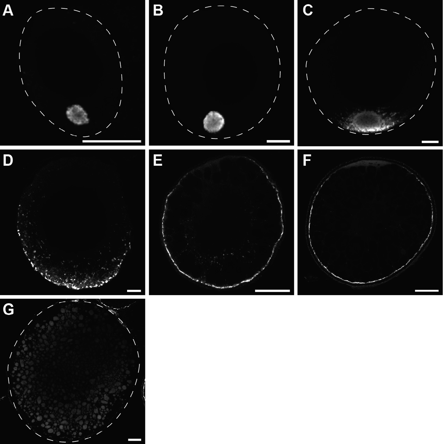
Fig. 2
Buc localization during oogenesis. Confocal images of wild-type oocytes, labeled with Buc antibody (white) at stage IA (A), early IB (B), late IB (C, D), early II (E), late II (F) and III (G). Buc moves with the germ plasm to the vegetal pole. Stippled line indicates outline of oocytes (A–C, G). Lateral views, animal to the top. Scale bar: 10 µm (A–D) and 50 µm (E–G).
Figure Data
Acknowledgments
This image is the copyrighted work of the attributed author or publisher, and
ZFIN has permission only to display this image to its users.
Additional permissions should be obtained from the applicable author or publisher of the image.
Reprinted from Gene expression patterns : GEP, 18(1-2), Riemer, S., Bontems, F., Krishnakumar, P., Gömann, J., Dosch, R., A functional Bucky ball-GFP transgene visualizes germ plasm in living zebrafish, 44-52, Copyright (2015) with permission from Elsevier. Full text @ Gene Expr. Patterns