
|
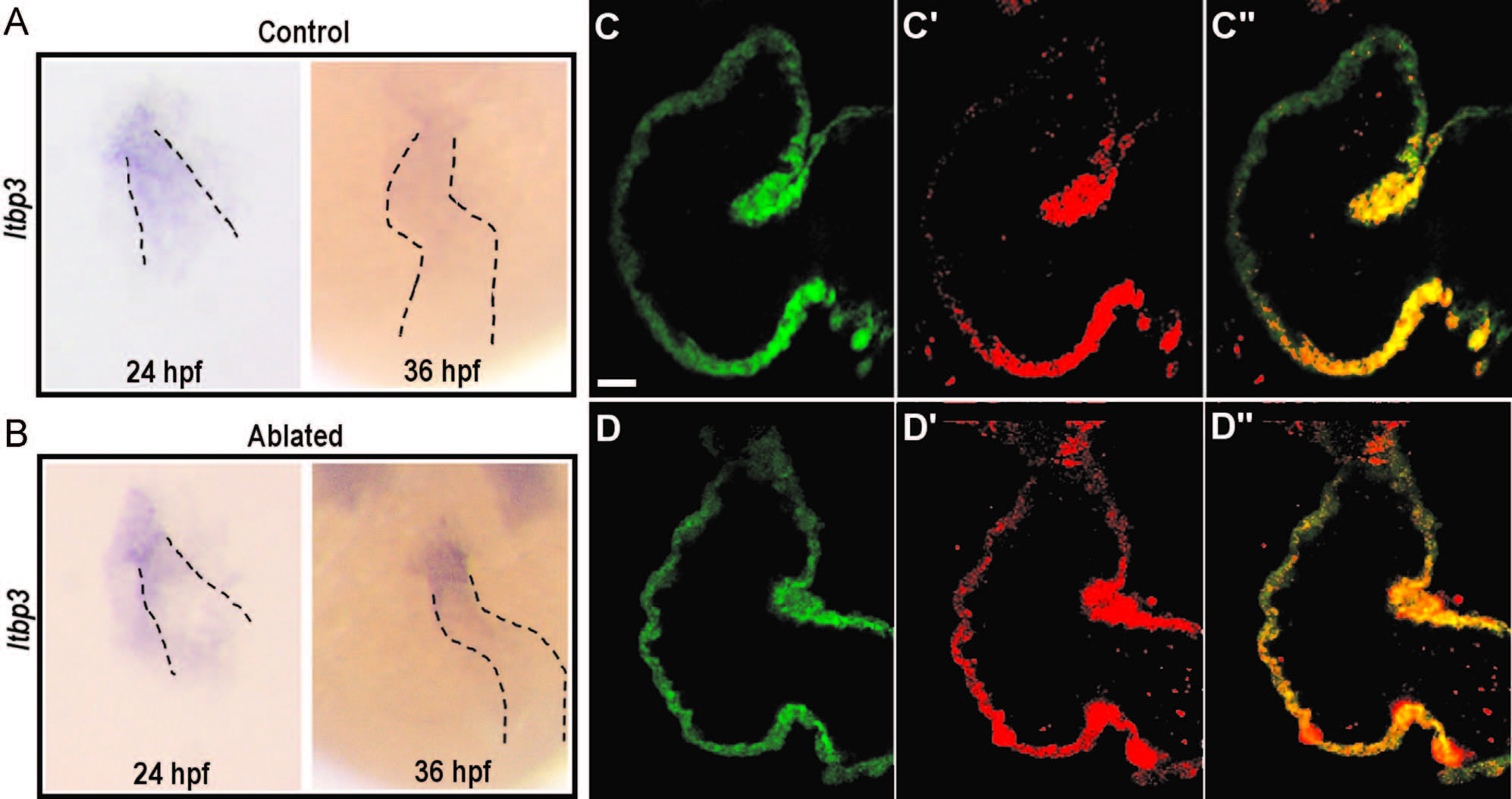
Fig. 7
Requirement for neural crest cells in the recruitment of second heart field progenitors. (A and B) In situ hybridization for ltbp3 in control (A) and NC-ablated embryos (B). Ltbp3 expression is observed in both control and NC-ablated embryos at 24 hpf. By 36 hpf, ltbp3 expression is down-regulated in control, but is persistent in NC-ablated embryos. Dashed lines indicate location of heart. (C–D′′) Single z sections of ventricles from Mtz-treated NfsB-mCherry (-); myl7:kaede control embryo (C–C′′) and Mtz-treated NfsB-mCherry (+); myl7:kaede NC-ablated embryos (D–D′′) at 60 hpf. Kaede protein photoconverted at 26 hpf are fluorescent in red whereas newly produced protein are in green. Note that ventricular cardiomyocytes at the arterial end express green Kaede whereas red Kaede proteins are present throughout the myocardium in NC-ablated embryos, indicating a defect in the recruitment of second heart field progenitors. Scale bar=20 µm.
Reprinted from Developmental Biology, 404(2), Cavanaugh, A.M., Huang, J., Chen, J.N., Two developmentally distinct populations of neural crest cells contribute to the zebrafish heart, 103-12, Copyright (2015) with permission from Elsevier. Full text @ Dev. Biol.