
|
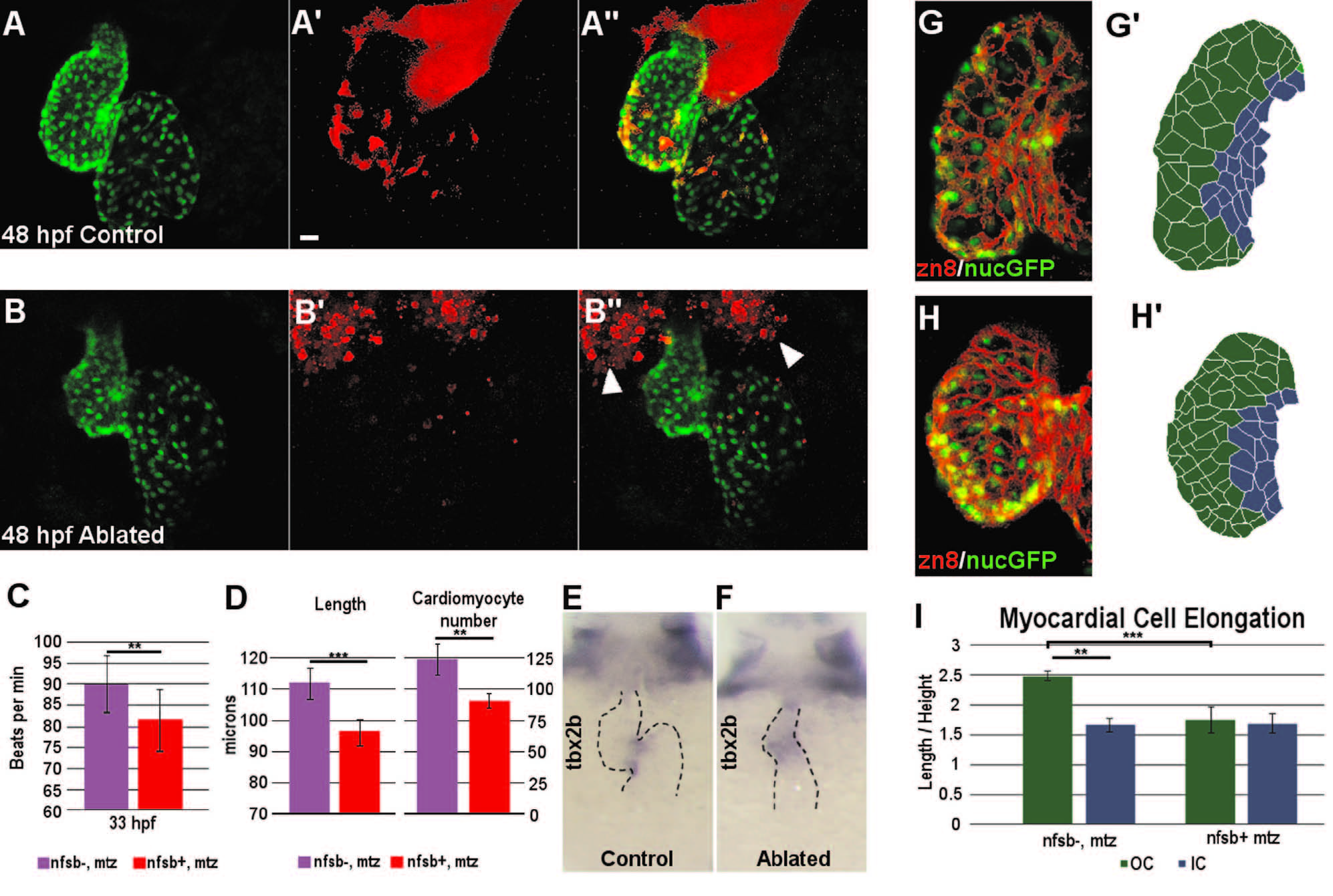
Fig. 6
Neural crest ablation affects cardiac function and morphology. (A–B′′) Confocal projection of hearts from untreated control (A–A′′) and Mtz-treated (B–B′′) NC:NfsB-mCherry; myl7:nucGFP embryos. Note that Mtz-treated embryos had smaller ventricles. Arrowheads point to dying NCCs in surrounding tissues. Scale bar=20 µm. (C) Graph indicates heart rate in Mtz-treated NfsB-mCherry (–) controls and Mtz-treated NfsB-mCherry (+) NC-ablated embryos at 33 hpf. (D) Graph indicates ventricle length of Mtz-treated NfsB-mCherry () controls and Mtz-treated NfsB-mCherry (+) NC-ablated embryos at 48 hpf and ventricular myocardial cell number at 72 hpf. (E, F) In situ hybridization for tbx2b at 60 hpf in control (E) and NC-ablated embryos (F). Note that tbx2b expression domain is expanded in NC-ablated embryos. (G, H) Confocal projections of 80 hpf ventricles immunostained with membrane marker Zn8 from control (G) and NC-ablated embryos (H). (G2, H2) Traces of cell shape from the images shown in G and H with outer curvature cells in green and inner curvature cells in blue. (I) Quantification of the ratio between the longest and shortest axis of each outer (green) and inner (blue) curvature myocardial cell in control and NC-ablated embryos. **p<0.05; ***p<0.001.
Reprinted from Developmental Biology, 404(2), Cavanaugh, A.M., Huang, J., Chen, J.N., Two developmentally distinct populations of neural crest cells contribute to the zebrafish heart, 103-12, Copyright (2015) with permission from Elsevier. Full text @ Dev. Biol.