
|
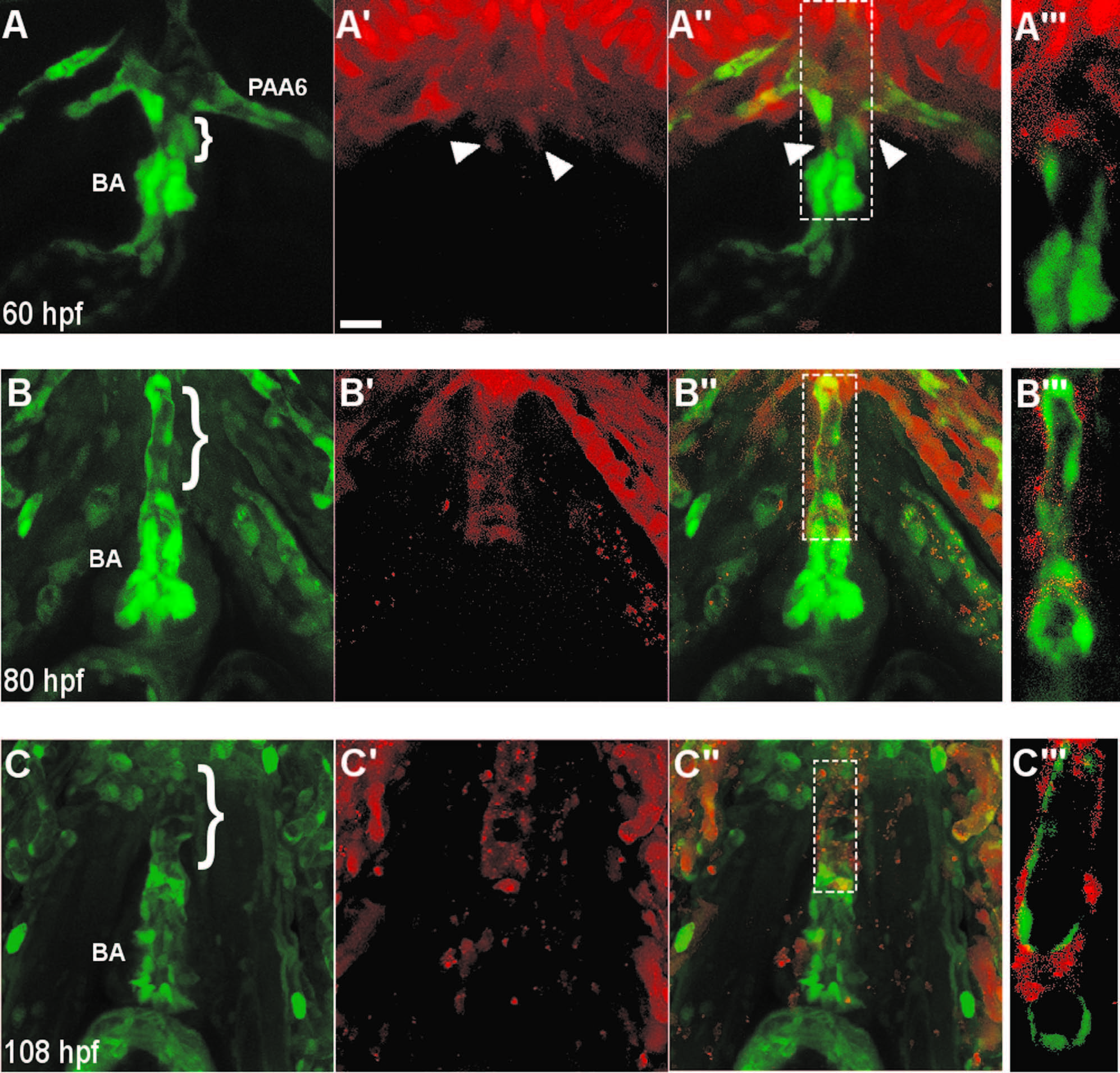
Fig. 4
Neural crest-derived cells surround the endothelium of the ventral aorta and bulbus arteriosus. (A–A′′) A confocal projection of a ventral view of the BA and VA of a NC:NfsB-mCherry; kdrl:GFP embryo at 60 hpf (anterior toward top). Arrowheads point to neural crest cells migrating onto the VA from the 6th aortic arch artery. (B–C3) Confocal projections of BA and VA of a NC:mCherry; kdrl:GFP embryo at 80 hpf (B–B′′′ and 108 hpf (C–C′′′) show the progression of mCherry+ cells migrating along the VA. A′′′, B′′′ and C′′′, Single z section of area boxed in A′′, B′′ and C′′, respectively. Brackets mark the ventral aorta (VA); BA, bulbus arteriosis; PAA6, pharyngeal arch artery 6, scale bar=20 µm.
Reprinted from Developmental Biology, 404(2), Cavanaugh, A.M., Huang, J., Chen, J.N., Two developmentally distinct populations of neural crest cells contribute to the zebrafish heart, 103-12, Copyright (2015) with permission from Elsevier. Full text @ Dev. Biol.