
|
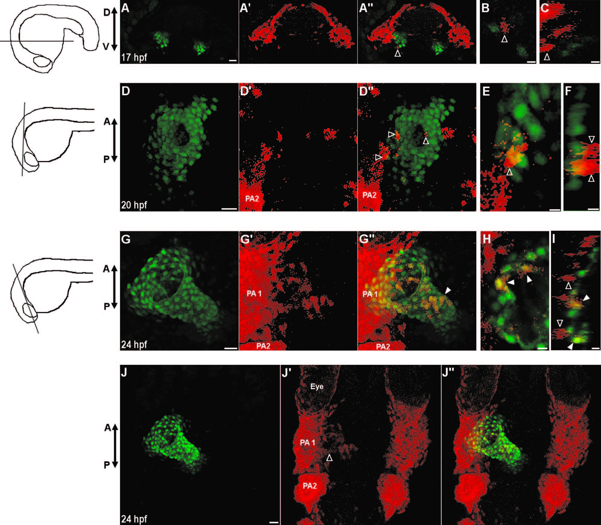
Fig. 2
Neural crest cells contribute to the primitive heart tube. (A–J) Confocal images of vibratome sections of NC:NfsB-mCherry; myl7:nucGFP embryos. Orientation of each section is indicated by illustration on the left. Sox10 positive cells are marked by mCherry and myl7 positive cardiomyocytes express nuclear GFP. At 17 hpf (A–C) and 20 hpf (D–F) mCherry+ cells are in close proximity of the heart and have not adopted a myocardial fate (open arrowhead). (G–I) At 24 hpf, some mCherry+ cells have integrated into the primitive heart tube and express nucGFP (closed arrowhead) while those mCherry+ cells adjacent to the heart do not (open arrowheads). (J–J′′) Dorsal view of the anterior region of the same embryo in G, showing mCherry+ cells migrating out of PA1 (open arrowhead). (A, D, G, J) Confocal projections of vibrotome sections, scale bar=20 µm. (B, E, H) Single confocal section, scale bar=5 µm. (C, F, I) Orthogonal reconstruction, scale bar=5 µm. PA1, pharyngeal arch 1; PA2, pharyngeal arch 2, D, dorsal; V, ventral; A, anterior; P, posterior.
Reprinted from Developmental Biology, 404(2), Cavanaugh, A.M., Huang, J., Chen, J.N., Two developmentally distinct populations of neural crest cells contribute to the zebrafish heart, 103-12, Copyright (2015) with permission from Elsevier. Full text @ Dev. Biol.