
|
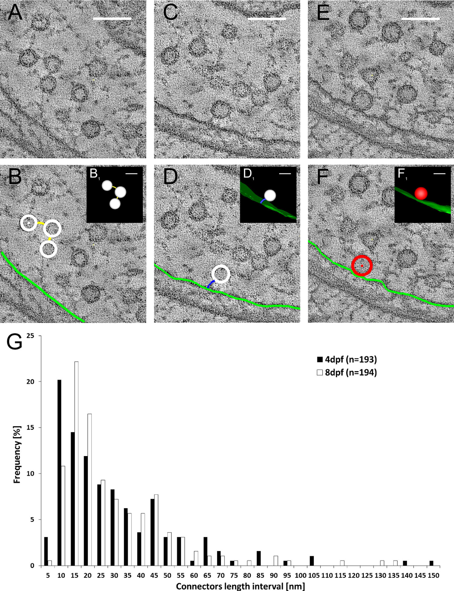
Fig. 5
The filamentous network between synaptic vesicles and between the membranes and vesicles. Single tomogram slices are shown, in which synaptic vesicles are interconnected by filaments (connectors; A,B), and connected to the membrane via small filaments (tethers; C,D). Example of a docked synaptic vesicle located directly at the cell membrane without a visible filament between is shown in E and F. In B, D, and F the synaptic vesicles are annotated in white, the connectors in yellow, the tethers in blue, the cell membrane in light green, and the docked vesicles in red. B1, D1, and F1 show 3D models of the annotated structures. The connectors between synaptic vesicles were measured in 4-dpf and 8-dpf zebrafish larvae (G). A magenta/green version of this figure is available as Supporting Information Figure 1. Scale bars = 100 nm in A-F; 50 nm in B1-F1.