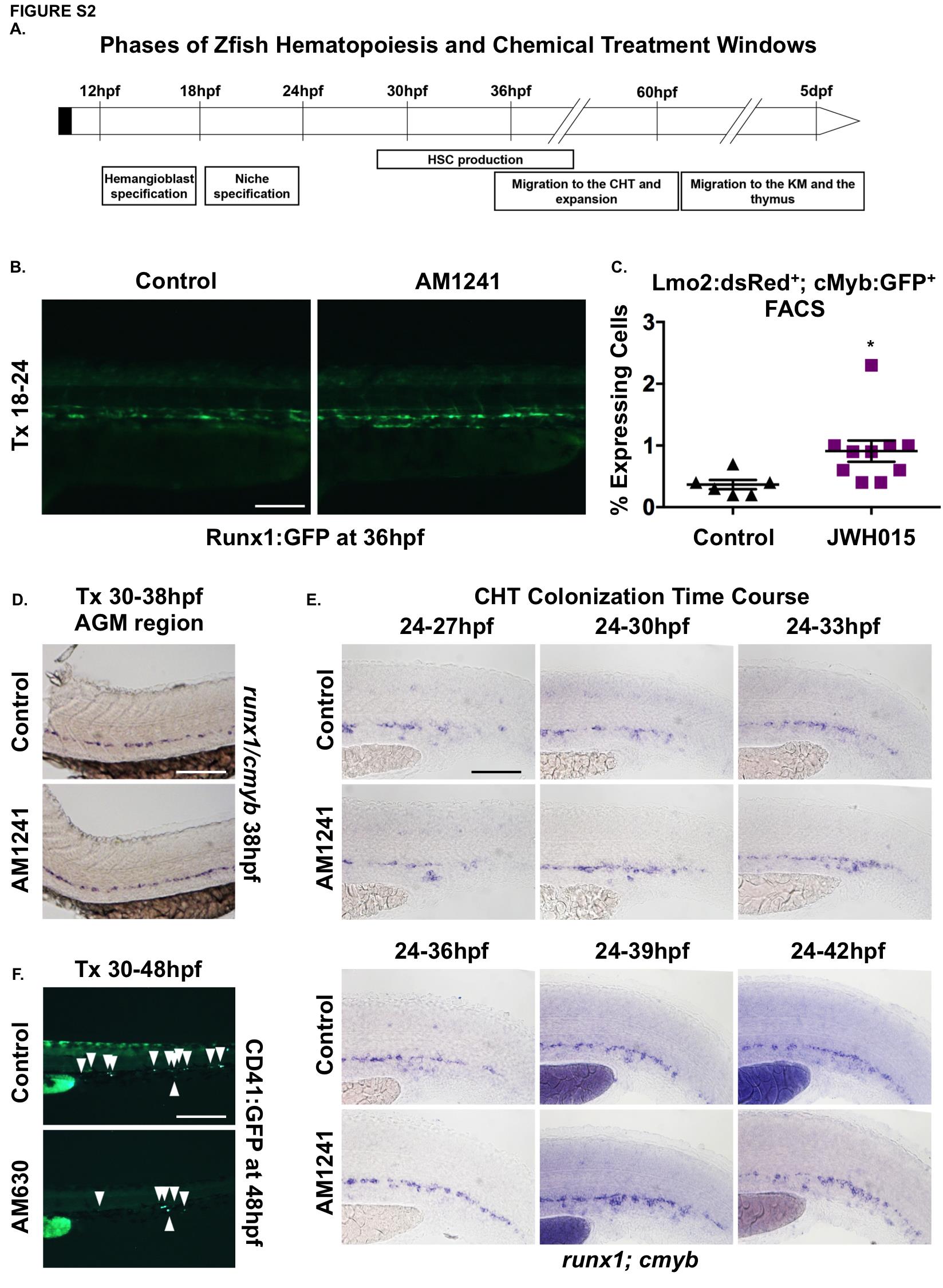
Fig. S2 CNR2-signaling is required for HSC production, expansion and CHT colonization.
(A) Schematic representation of the different stages of HSC development starting during somitogenesis (12hpf) and continuing until larval stages (5dpf). HSCs are specified in the hemogenic endothelium prior to 24hpf, and are actively produced between 28 and 48hpf in the AGM. HSCs bud and migrate to CHT starting around 33hpf. HSC expansion occurs in the CHT, and HSCs begin to exit to seed the kidney marrow (KM) and thymus by 72hpf.
(B) In vivo imaging confirmed numbers of Runx1:GFP+ HSPCs were increased in the AGM at 36hpf following AM1241 exposure during niche specification (18-24hpf) (n≥10/condition).
(C) The effect of JWH015 (18-24hpf) on HSPCs was quantified by FACS analysis of cmyb:gfp;lmo2:dsRed2 embryos (2.48-fold; *p<0.05, 2-tailed t-test, n≥6/condition). (D) Embryos exposed to AM1241 during HSC production (30-38hpf) exhibited no apparent increase in runx1;cmyb+ HSPCs within the AGM region (n≥65/condition).
(E) Time course analysis (24-42hpf) of the qualitative phenotypic distribution of runx1;cmyb expression in the CHT, indicating increased HSPC colonization in response to AM1241-treatment as compared to controls (ne55/condition).
(F) In vivo imaging of cd41:egfp embryos indicated the number of CD41:GFP+ HSCs (arrowheads) was decreased in the CHT at 48hpf following exposure to AM630 (n≥15/condition; see also Figure 2K).
Scale bars: B,D,E,F=100µm.

