
|
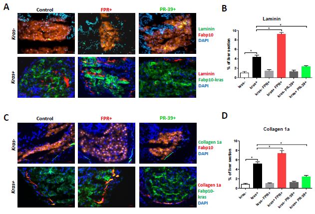
Fig. S2 Effect of neutrophil infiltration on expression of Laminin and collagen I in the liver. 8-dpf fabp10+ and kras+ larvae after exposure to FPR-A14 or PR-39 with or without doxycycline were cryo-sectioned and immune-stained for Laminin and Collagen I expression. (A, B) Immuno-staining for Laminin (A) and quantification (B). (C, D) Immuno-staining for Collagen 1a (C) and quantification (D). Control fabp10+ larvae with DsRed expression in hepatocytes were stained with Alexa Fluor 488-conjugated secondary antibody after the primary antibody incubation (top rows) while kras+ larvae with GFP expression in hepatocytes were stained with Alexa Fluor 568-conjugated secondary antibody (bottom rows). All sections were counter-stained with DAPI. Treatment groups (n=10 each group) and color probes are indicated at the top and on the right respectively. Quantification of percentage of Laminin- or collagen I-positive areas out of total liver area is presented in (C) and (D). Statistical significance in quantification: *p<0.05.
Reprinted from Journal of hepatology, 63(2), Yan, C., Huo, X., Wang, S., Feng, Y., Gong, Z., Stimulation of hepatocarcinogenesis by neutrophils upon induction of oncogenic kras expression in transgenic zebrafish, 420-8, Copyright (2015) with permission from Elsevier. Full text @ J. Hepatol.