
Fig. 8
- ID
- ZDB-IMAGE-150923-14
- Genes
- Antibodies
- Publication
- Yan et al., 2015 - Stimulation of hepatocarcinogenesis by neutrophils upon induction of oncogenic kras expression in transgenic zebrafish
- All Figures
- Figures for Yan et al., 2015
|
Fig. 8
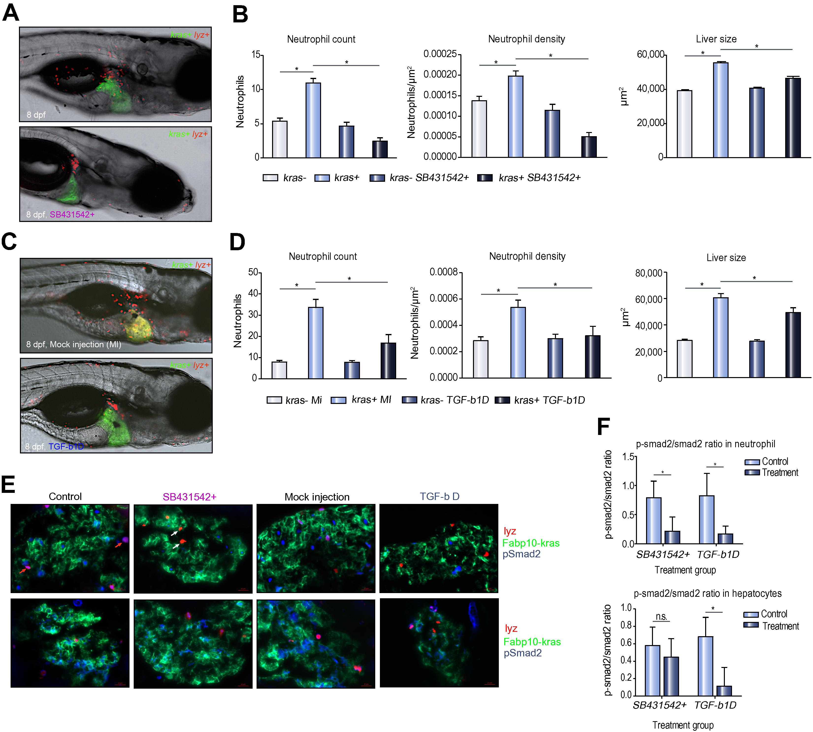
Effect of antibody-mediated Tgf-β depletion on TAN recruitment. (A) Representative images of 8 dpf kras+/lyz+ larvae exposed to either doxycycline alone (top) or with SB431542 (bottom). (B) Neutrophil counts (left) and density (middle) in the liver and liver size (right) after SB431542 treatment. (C) Representative images of 8 dpf kras+/lyz+ larvae injected with Tgf-β antibody (bottom) or mock injected as a control (top). Both were induced by doxycycline. (D) Neutrophil counts (left) and density (middle) in the liver and liver size (right) in Tgf-β-depleted (tgfb-1D) and mock injected (MI) larvae. (E) Validation of SB431542 exposure and Tgf-β depletion by immunostaining of Smad2 and phospho-Smad2 (pSmad2). Representative liver sections are shown from each group as indicated. The color code of each probes correspond the color signals in the images. Red arrows, DsRed expressing neutrophils with pSmad2 expression; white arrows, DsRed expressing neutrophils without pSmad2 expression. (F) Ratios of pSmad2/smad2 in neutrophils (top) and hepatocytes (bottom) in SB431542-inhibited and Tgf-β depleted larvae. n = 10; *p <0.05.
Reprinted from Journal of hepatology, 63(2), Yan, C., Huo, X., Wang, S., Feng, Y., Gong, Z., Stimulation of hepatocarcinogenesis by neutrophils upon induction of oncogenic kras expression in transgenic zebrafish, 420-8, Copyright (2015) with permission from Elsevier. Full text @ J. Hepatol.