
Image
|
Figure Caption
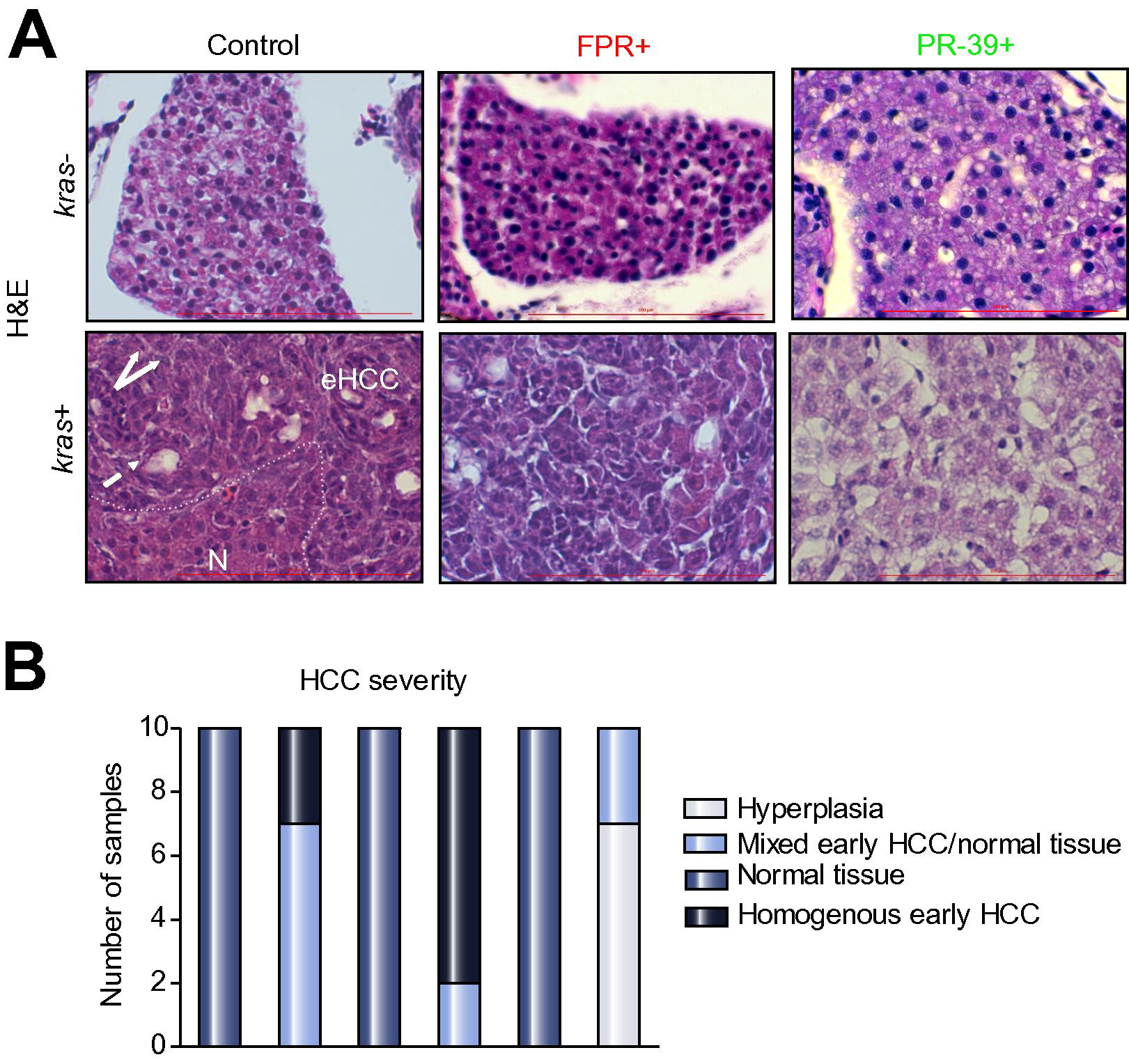
Fig. 6
Histological analyses of effects of neutrophils on hepatocarcinogenesis. (A) H&E staining of liver sections. 8 dpf kras-/lyz+ and kras+/lyz+ larvae after exposure to FPR A14 or PR-39 with or without doxycycline, as indicated in the pictures, and were subject to H&E staining. Abbreviations: eHCC, early HCC; N, normal liver region. Dashed arrow: pseudo glandular pattern; white arrows: enlarged nuclei. (B) Quantification of phenotype observed in liver histology sections (n = 10, *p <0.05).
Figure Data
Acknowledgments
This image is the copyrighted work of the attributed author or publisher, and
ZFIN has permission only to display this image to its users.
Additional permissions should be obtained from the applicable author or publisher of the image.
Reprinted from Journal of hepatology, 63(2), Yan, C., Huo, X., Wang, S., Feng, Y., Gong, Z., Stimulation of hepatocarcinogenesis by neutrophils upon induction of oncogenic kras expression in transgenic zebrafish, 420-8, Copyright (2015) with permission from Elsevier. Full text @ J. Hepatol.