
|
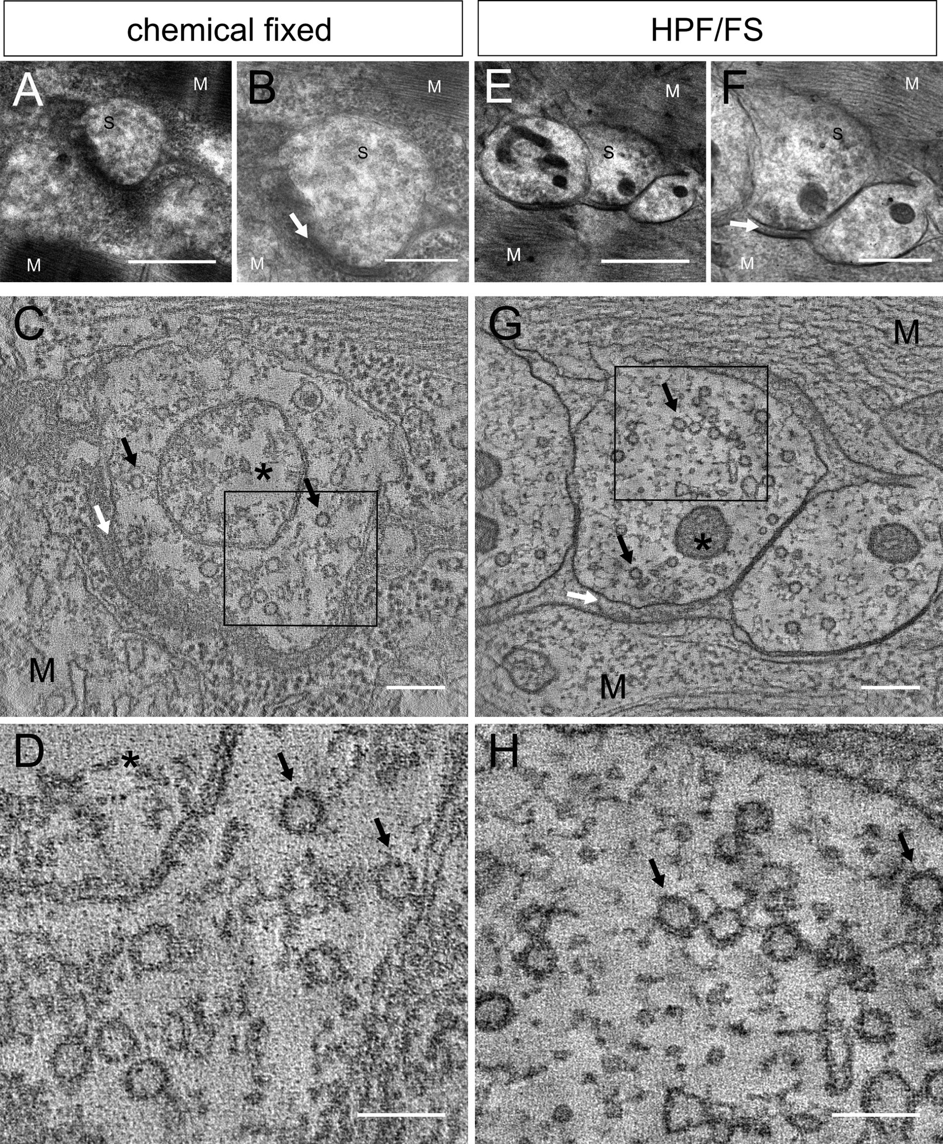
Fig. 1
Differences in ultrastructural preservation in chemically fixed and cryoimmobilized and freeze substituted samples. The overview shows electron micrographs of a chemically fixed (A) and an HPF/FS-treated (E) synapse (S) of a ~250-nm-thick section from a 4-dpf zebrafish larva. Synapses are surrounded by muscle cells (M) with an electron-dense synaptic cleft (white arrows) in between (B,C,F,G). C and G show a tomogram slice per synapse. Mitochondria (asterisks) and synaptic vesicles (black arrows) are annotated. A higher magnification image of the boxed region in C and G is shown in D and H, respectively. Scale bars = 1 µm A,E; 500 nm in B,F; 200 nm in C,G; 100 nm in D,H.