
|
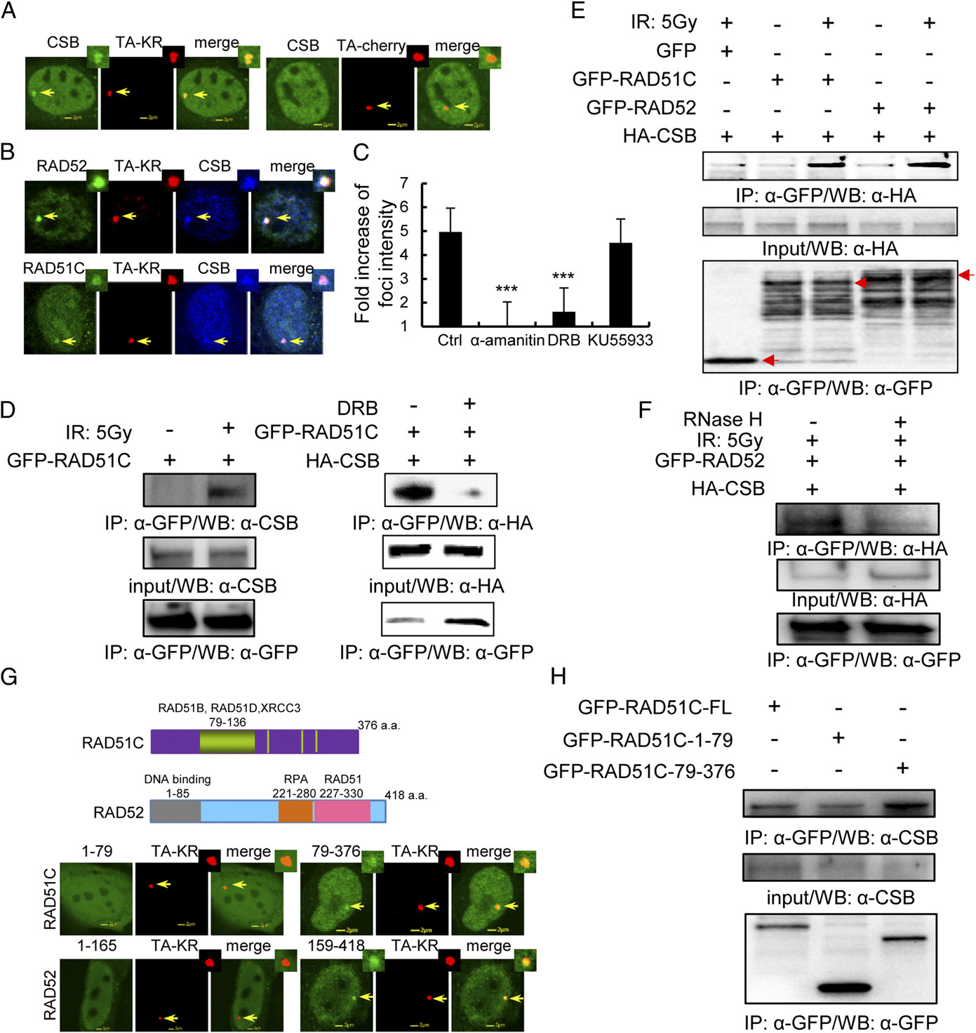
Fig. 4
CSB localizes at TA-KR damage sites and interacts with RAD52 and RAD51C after DNA damage. (A) Recruitment of CSB at sites of TA-KR after damage. FLAG-CSB and TA-cherry/KR were cotransfected into U2OS TRE cells. Twenty-four hours later, cells were irradiated with light for 10 min followed by 30 min of incubation; the recruitment of CSB at TA-KR sites is shown with FLAG-antibody. (B) Colocalization of CSB, RAD52, or RAD51C at sites of TA-KR after damage. FLAG-CSB, GFP-RAD52/RAD51C, and TA-KR were cotransfected into U2OS TRE cells. Twenty-four hours later, cells were irradiated with light for 10 min followed by 30 min of incubation and stained with FLAG-antibody. (C) The average fold increase of FLAG-CSB at sites of TA-KR after damage (30 min postlight incubation after 10 min of light incubation) and after treatment with α-amanitin (100 µg/mL) for 1 h, DRB (20 µM) for 24 h, or KU55933 (10 µM) for 1 h. Images were quantified by ImageJ (n > 50); SEM indicates three independent experiments. (D and E) GFP-RAD52/GFP-RAD51C stably expressed in 293 cells with or without 5 Gy IR and DRB treatment was analyzed by IP with anti-GFP and 1 h postirradiation incubation. Detection of endogenous CSB or HA-tagged CSB is shown. (F) RNaseH treatment of cells abolishes the interaction between CSB and RAD52. HA-CSB expressed in GFP-RAD52 stably expressing 293 cells treated with 5 Gy IR, analyzed by IP with anti-GFP and 1 h postirradiation incubation with or without RNaseH (15 units) treatment for 15 min. Detection of HA-tagged CSB is shown. (G) The recruitment of the C-terminus RAD52 and RAD51C at TA-KR 30 min after KR activation. (H) CSB interacts with the C terminus of RAD51C. GFP-RAD51C fragments were transiently transfected into 293 cells. Forty-eight hours after transfection, cells were irradiated with 5 Gy IR. Cell lysates were collected 1 h after irradiation and immunoprecipated by GFP/HA antibody. Western blotting by anti-HA, anti-GFP, and anti-CSB for immunoprecipitates and input are shown.