
IMAGE
Fig. S3
- ID
- ZDB-IMAGE-150916-23
- Publication
- Simões et al., 2014 - Denervation impairs regeneration of amputated zebrafish fins
- All Figures
- Figures for Simões et al., 2014
Image
|
Figure Caption
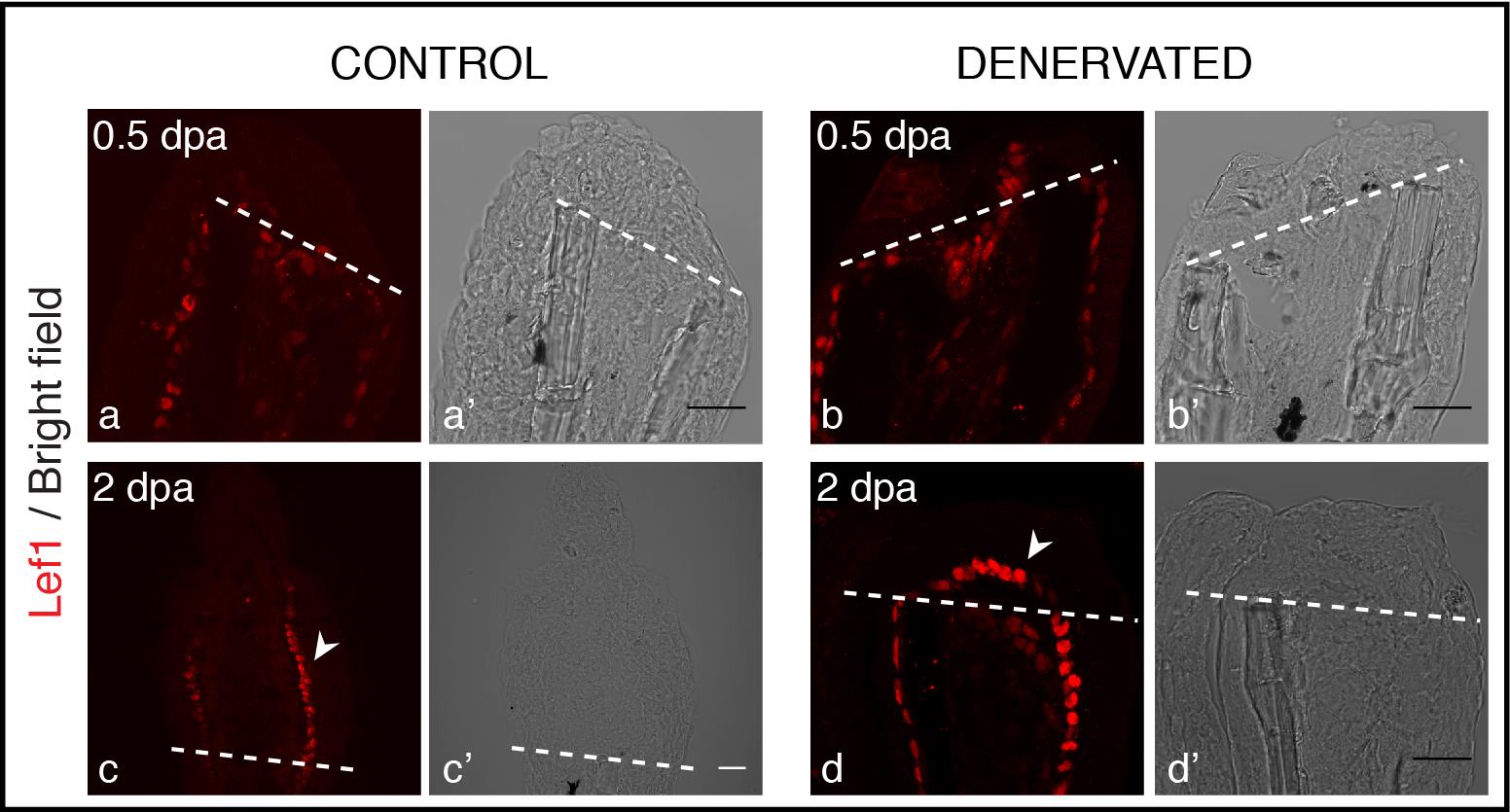
Fig. S3
Staining for Lef1 in longitudinal sections after amputation of denervated fins. Lef1 protein is detected in the BEL of both control and denervated fins from 0.5 to 2 dpa. At 0.5 dpa Lef1 expression is similar in control (a) and denervated (b) fins. At 2 dpa Lef1 is restricted to the most proximal BEL cells in control fins (c-arrowhead) and it is expressed all over the BEL lining mesenchymal cells of denervated fins (d-arrowhead). Scale bar - 25 µm. a-d) The images are a projection of confocal optical slices. Dashed lines mark amputation plane. Scale bar - 25 µm.
Acknowledgments
This image is the copyrighted work of the attributed author or publisher, and
ZFIN has permission only to display this image to its users.
Additional permissions should be obtained from the applicable author or publisher of the image.
Full text @ BMC Dev. Biol.