
|
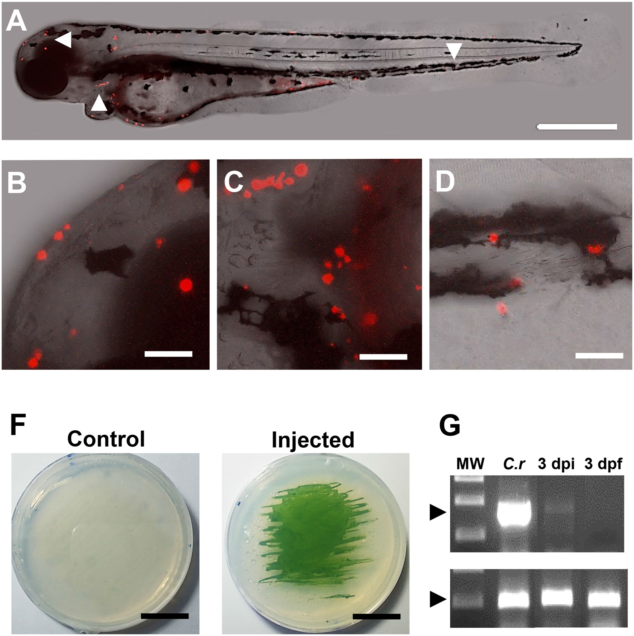
Fig. 4 Distribution and viability of microalgae in the zebrafish larvae.
C. reinhardtii was microinjected and visualized at 3 dpf. Results shows that algae distribute along the whole larva (A), including anterior (B), meddle (C) and posterior areas (D). At 3 days post fertilization (3 dpi), injected or control embryos were disaggregated and placed in agar plates, showing the capacity of the alga to re-growth ex-vivo (F). RT-PCR shows the expression of the alga specific gene psbD in RNA extracts obtained from C. reinhardtii (C.r) and fishes at 3 days post injection (3 dpi). No signal was detected in the non-injected fish at 3 days post (3dpf; D). Scale bar represents 500 µm in A and 50 µm in B-D and 1.5 cm in F. Arrow heads in A indicate the areas shown in B-D. n ≥ 5 in A-D and n = 3 in F and G.