
|
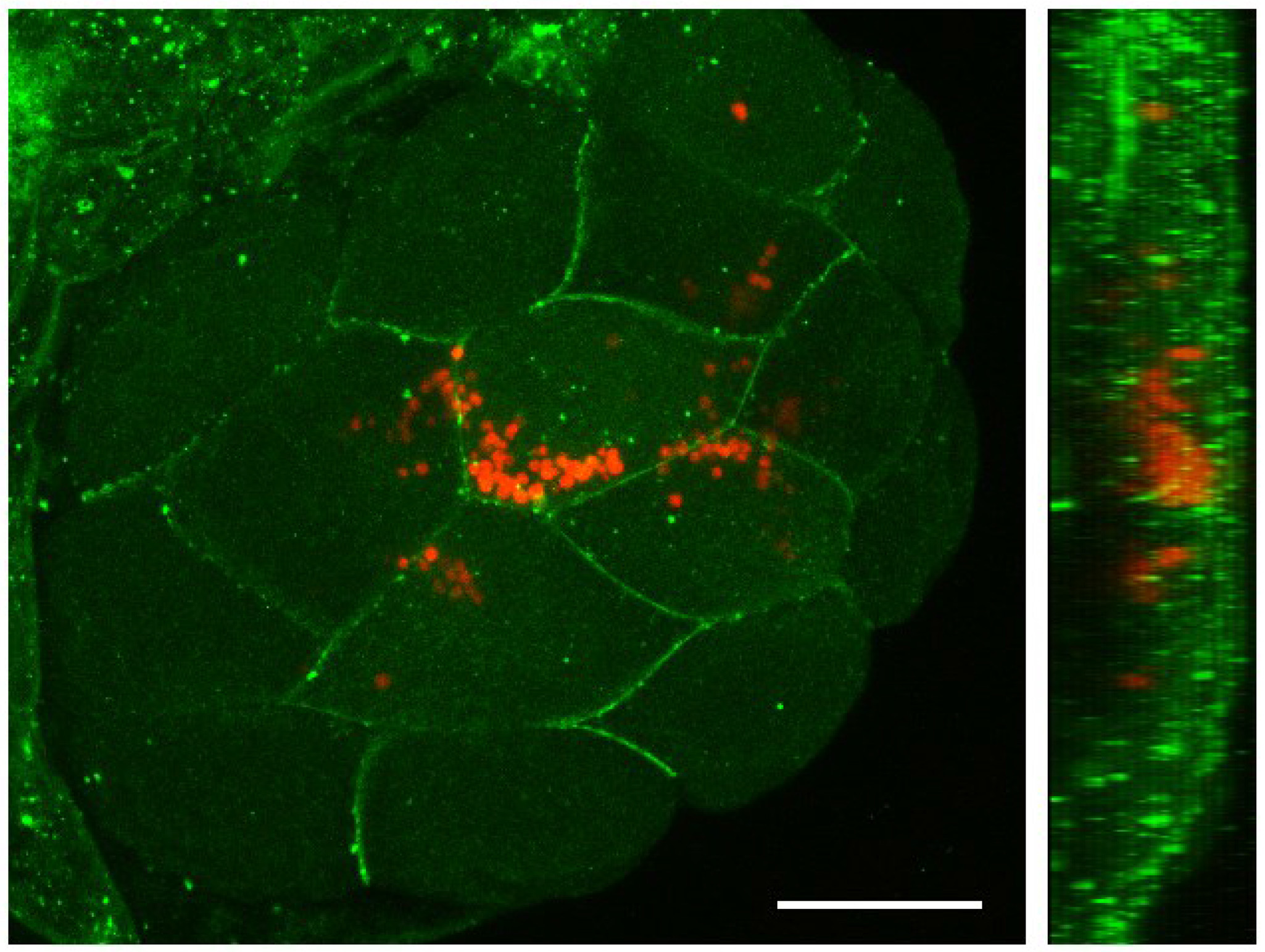
Fig. 3 Distribution of Chlamydomonas reinhardtii in early zebrafish embryos.
A zebrafish embryo at one cell stage was injected with a suspension of algae, raised to the 16 cell stage (1.5 hpf) and processed for immunohistochemistry. The blastoderm was imaged under confocal microscopy to reveal that microalgae were mainly located intracellularly. Cell membranes stained with anti-β-catenin antibody are shown in green, while C. reinhardtii is observed in red (autofluorescence). A Z-stack projection is shown on the left and a reconstructed Y projection view of the same embryo is observed on the right. Scale bar represents 200 µm. n ≥ 10.