
|
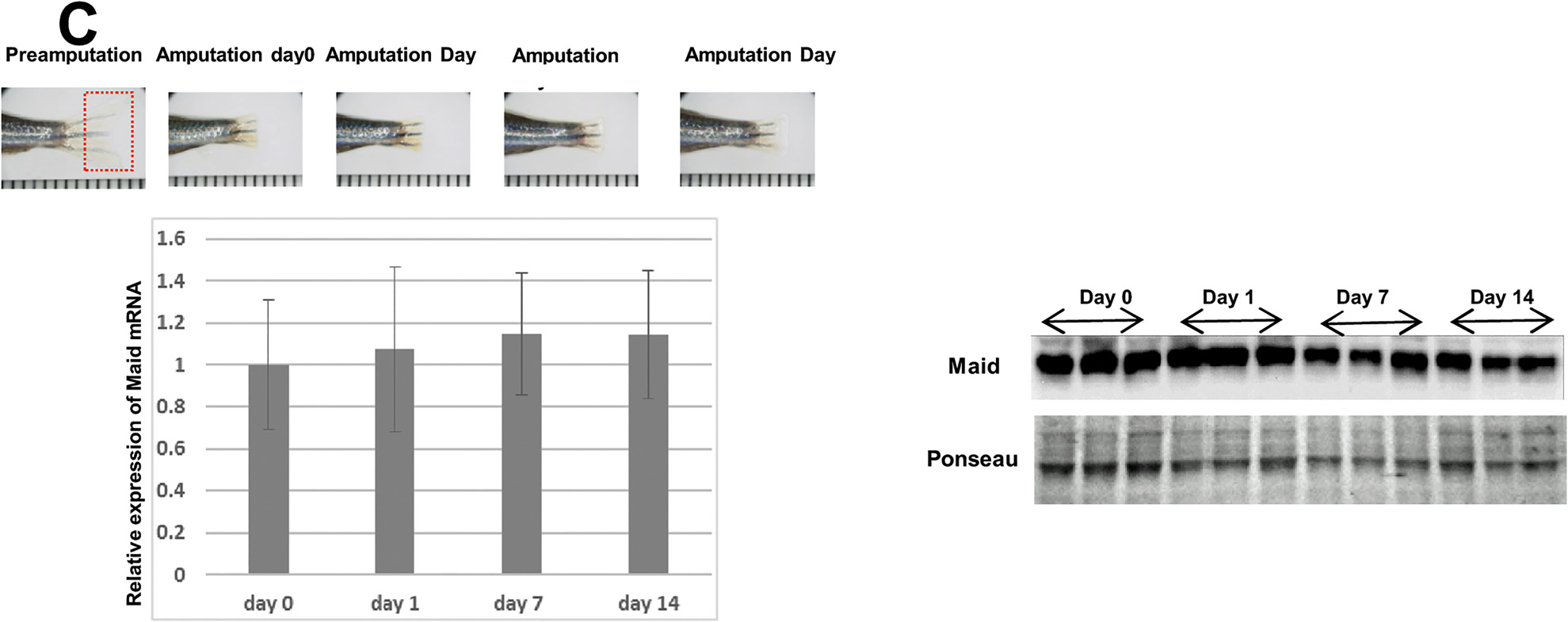
Fig. 3 Maid expression following partial hepatectomy or fin amputation in zebrafish.
(A) Top: Diagram of the area of adult zebrafish liver subjected to PH. Middle: Macroscopic views of fish abdomens, sliced in the middle. About 30% of the ventral lobe of the liver was removed. The area of the liver regeneration is demarcated by a rectangle. Bottom: Macroscopic view of the livers in the middle panel after 7 days of regeneration. Results are representative of 10 fish per group. (B) Left: RT-PCR analysis of Maid mRNA expression in regenerated zebrafish liver at the indicated times after PH. Results are the mean ± SD expressed relative to Gapdh and are representative of at least 5 fish per group, *, p<0.05. Right: Western blotting analysis of Maid in regenerated zebrafish liver. (C) Left Top: Macroscopic views of a representative adult zebrafish tail fin before and after amputation. Fin regeneration was monitored for 14 days. Left Bottom: RT-PCR analysis of Maid mRNA expression in regenerating fin at the indicated times after fin amputation. Results are the mean ± SD (n = 3/group) and are expressed relative to El1a mRNA in fish at Pre-Amputation. Right: Western blotting analysis of Maid in regenerating fin.