
|
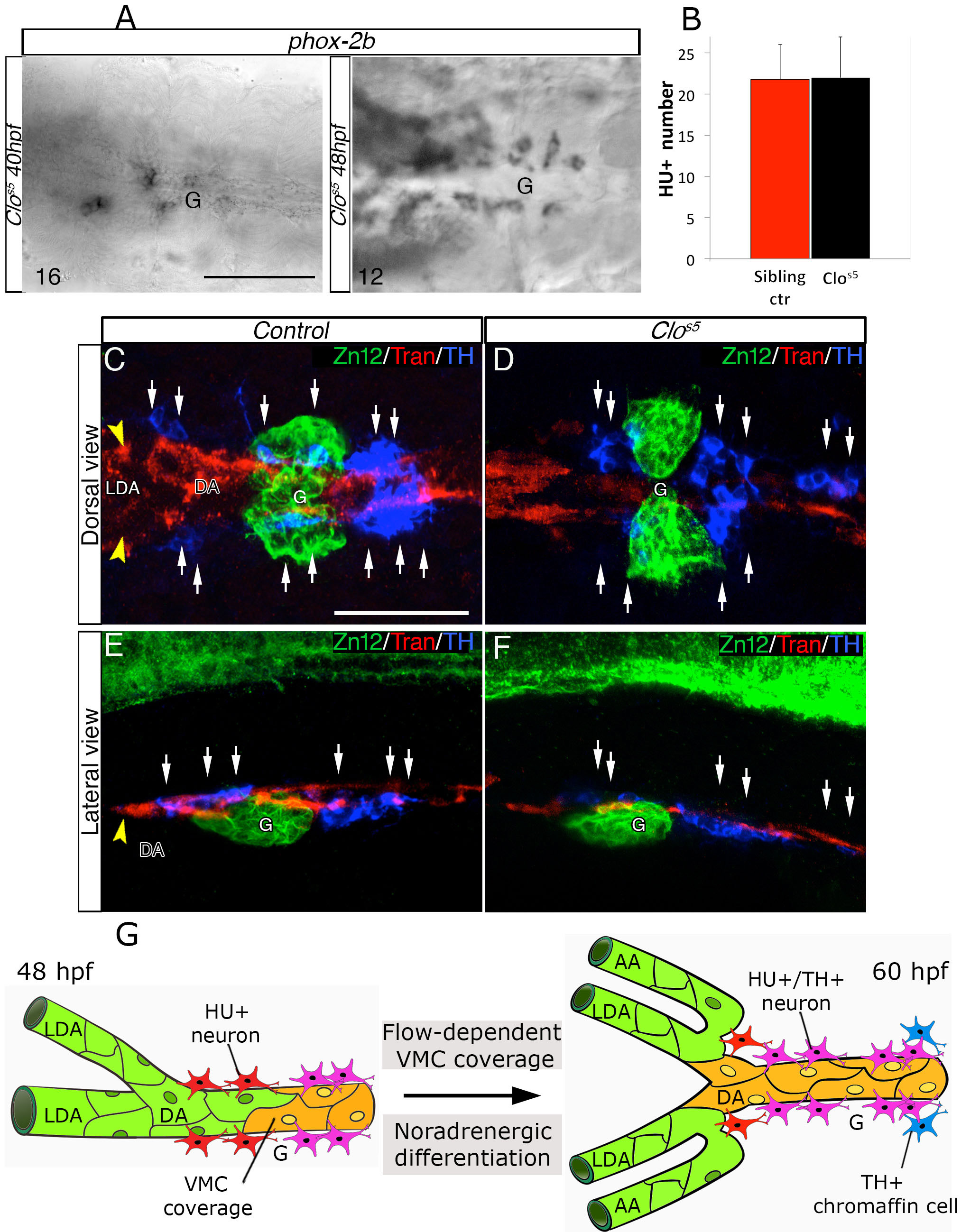
Fig. S6 SN differentiation in clos5 mutants, related to Figure 7. (A) Phox-2b RNA expression was analyzed in clos5 Tg(kdrl:EGFP) embryos by in situ hybridizations at the indicated developmental stages. Images are ventral views (anterior is to the left) around the glomerular region (G) after dissection of the yolk sac. Phox-2b positive sympathetic precursors are present around the DA at 40 and 48 hpf. The number of embryos observed is indicated at the bottom left. (B) Quantitative analysis of HU+ cells in Tg(kdrl:EGFP) control embryos and clos5 littermates at 60 hpf. Data were calculated from three independent experiments, error bars indicate SD. (C-F) Confocal analysis of control- (C, E) or clos5 (D, F) Tg(kdrl:EGFP) embryos. Dorsal (C, D) and lateral (E, F) views of the region around the glomerulus, anterior is to the left. Embryos are immunostained with antibodies to detect ZN12 (glomerulus, green), Transgelin (Tran) (red) and TH (blue). Note that SNs (white arrows) are present in proximity to VMCs in control and clos5 littermates. Scale bars: 75 µm in A-E. (G) Model for DA-mediated NA differentiation of SNs. Between 48 – 60 hpf, arterial vessels (LDA-DA connection) undergo angiogenic remodeling. Blood flow and PDGFR signaling enhance VMCs (yellow) recruitment around remodeling vessels. HU+ post-mitotic neurons (red) differentiate into NA neurons (HU+/TH+) (pink) in parallel with VMC differentiation. Fully differentiated SNs are detected in proximity to newly VMC-covered arterial vessels at 60hpf. DA, dorsal aorta; LDA, lateral dorsal aorta; AA, aortic arch; G, glomerulus; VMC, Vascular mural cells.