
|
Fig. 1
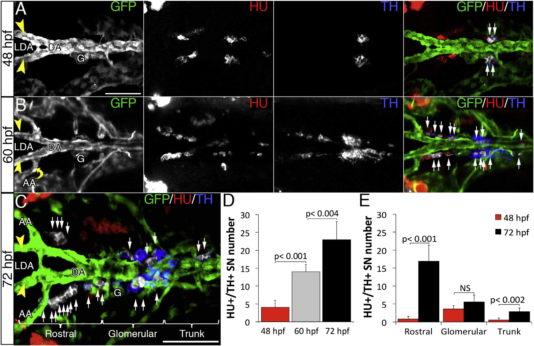
SNs Develop Next to the DA in Zebrafish Embryos
(A–C) Dorsal views (anterior is to the left) of whole-mount Tg(kdrl:EGFP)la116 zebrafish embryos between the LDA (arrowheads) and DA connection and the glomerular region (G) at the indicated time points. Immunostaining with antibodies to detect GFP (vessels, green), Hu C/D (post-mitotic neurons, red), and tyrosine hydroxylase enzyme (TH) (catecholaminergic marker, blue). (A and B) HU+/TH+ SNs are pseudocolored in white (arrows) in the merged confocal images. (C) Brackets mark the three clusters (rostral, glomerular, and trunk) of SNs organized around DA.
(D) Quantification of total number of double HU+/TH+ cells at 48, 60, and 72 hpf.
(E) Quantification of double HU+/TH+ cells organized in three clusters around the DA at 48 and 72 hpf. Data were calculated from three independent experiments. NS, not significant; error bars indicate SD.
DA, dorsal aorta; LDA, lateral dorsal aorta; AA, aortic arch; G, glomerulus; hpf, hours post-fertilization. Scale bars, 75 µm (A–C). See also Figure S1>.